China Wholesale Digital Dermoscopy Factory Manufacturer Supplier - IBOOLO

Digital Marketing, Defined by Mailchimp, Is A Concept That Encompasses Various Online Strategies And Tactics for Businesses to Effectively Promote Their Products Or Services in The Digital Realm.

The practice of digital marketing, also known as online marketing, involves the endorsement of brands through the utilization of the internet and other digital means to establish connections with prospective customers. This encompasses a broad spectrum of channels, such as email, social media, web-based advertising, as well as text and multimedia messages, effectively serving as a comprehensive marketing strategy.

What Does A Digital Marketing Agency Do?

A digital marketing agency is responsible for executing a wide range of tasks to ensure success in the digital marketing realm. Whether it's developing effective strategies, executing campaigns, or optimizing online presence, these agencies handle it all. Their extensive services cover various areas such as search engine optimization, social media management, content creation, email marketing, and pay-per-click advertising. They meticulously analyze market trends and consumer behavior to tailor their tactics for maximum impact. In essence, a digital marketing agency acts as a one-stop solution for businesses aiming to thrive in the digital landscape.

With its omni-channel, multi-channel, or single-channel approach, a digital marketing agency effectively connects with customers in the online realm. Through various channels like websites, blogs, email, and social media platforms, agencies engage with customers and ensure a comprehensive interaction experience.

People May Ask

Which Skin Cancer Type Poses A Greater Threat: Basal Cell Carcinoma Or Squamous Cell Carcinoma?

While therapeutic interventions achieve positive outcomes in approximately 90% of instances involving both cancer types, namely basal cell carcinoma and squamous cell carcinoma, it is noteworthy that for certain individuals, the latter condition can pose a more severe health threat. Amongst these two, squamous cell carcinoma exhibits a heightened tendency to disseminate to other bodily regions, albeit such occurrences are infrequent.

What Is The Price Range for Undergoing A Visia Skin Analysis?

The typical expenditure for undergoing a VISIA® skin assessment at the Belladerma Cosmetic Surgery & Skin Care Center approximates to $150.

What Is The Visual Representation of Dermoscopy?

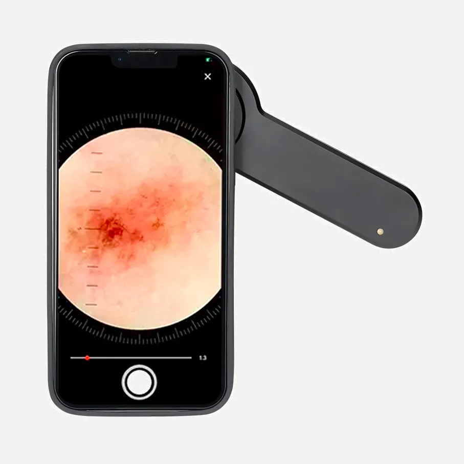
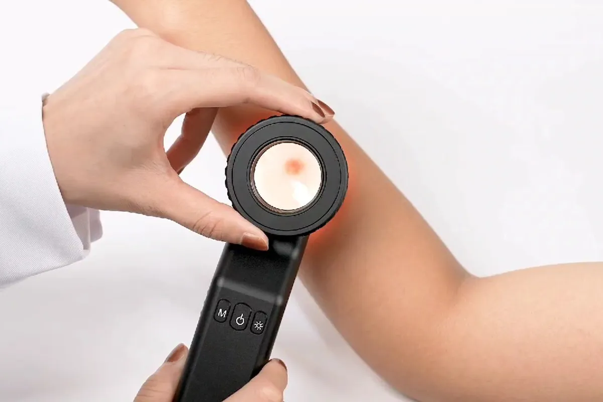
A dermatoscope possesses the capability to evaluate the structures extending down to the depth of the reticular dermis, allowing for the capture of images for subsequent comparison. The fundamental concept behind dermoscopy involves transilluminating a lesion, enabling detailed examination through the use of high magnification to reveal intricate characteristics.

Can Blood Tests Reveal The Presence of Skin Cancer?

The presence of skin cancer cannot be identified through a comprehensive blood examination or any alternative blood analysis. However, once a diagnosis of skin cancer has been made, blood tests may serve as a tool to ascertain the extent of the disease or evaluate the success of the prescribed treatments.

What Is The Price Range for Undergoing A Visia Scan?

The VISIA® skin assessment serves as a robust basis for gaining valuable insights, directing your personalized treatment strategy, and assessing the success of any administered therapies. What is the Typical Expense of Undergoing a VISIA® Skin Evaluation? At Belladerma Cosmetic Surgery & Skin Care Center, the average price tag for a VISIA® skin analysis amounts to $150.

Could You Elaborate on The Fundamental Concept behind Dermoscopy?

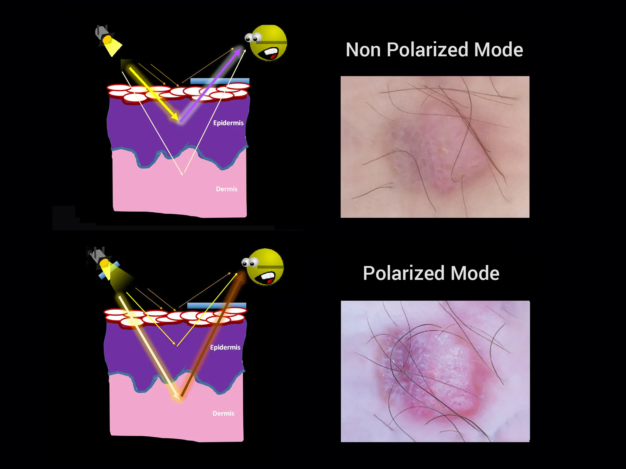
Dermatoscopy's core concept revolves around transilluminating a lesion to scrutinize it under high magnification, enabling the visualization of intricate details. Light encountering a surface, such as the skin, undergoes various interactions like reflection, refraction, diffraction, and/or absorption [Figure 1A]. These phenomena are shaped by the inherent physical attributes of the skin.

What Are The Distinguishing Factors between Confocal Microscopy And Laser Scanning Techniques?

A confocal setup employs laser illumination to concentrate on a minute area (or plane) within a sample, utilizing a tiny aperture to block out-of-focus light originating from the sample. The scanning of the laser involves guiding the laser beam through two mirrors in the Y and X directions to a precise spot on the sample.

What Are The Advantages of Utilizing Confocal Microscopy?

The primary advantage that confocal microscopy holds over alternative microscopy techniques lies in its capacity to restore the three-dimensional structure of specimens. This is achieved by scanning numerous planes and subsequently integrating them using dedicated deconvolution software, resulting in the reconstruction of 3D images.

What Is The Level of Efficacy Exhibited by Dermoscopy?

Dermoscopy sensitivity has been documented to vary between 60% and 100%, contingent upon various factors, including the proficiency of the inspecting clinicians and the complexity of the lesions under examination. While dermoscopy undoubtedly enhances melanoma diagnosis precision, it remains insufficient as a standalone diagnostic tool, necessitating histopathological analysis.

What Does The Term "Digital Mole Mapping" Refer to?

The utilization of digital mole mapping presents a secure and non-intrusive approach for skin cancer detection. This methodology incorporates a sophisticated digital photography system designed to chart the entirety of a person's moles. Subsequently, the captured images can be analyzed for any changes or the emergence of new moles over a period of time.

Digital Dermoscopy Products

Marketing Strategies: Lucrative Approaches in the Era of Digitalization - 9th Edition, Available as a Kindle Book

An Introduction to Digital Marketing Strategies in the Pharmaceutical Sector Softcover Edition – June 5th, 2019

Expand Your Business Horizons Through Video Marketing: Leverage the Power of Video Content with These Seven Tried-and-True Video Script Templates! Available in Kindle Edition

Introduction to Selling Digital Items on Etsy for Newbies (Home-Based Business Manual) Paperback – Released on March 24th, 2023

Creating a Profitable and Sustainable Digital Marketing Agency through the Establishment of a Successful Micro-Agency: A Guide [Copy Reproduction] Kindle Version

The Simple English Guide to SEO: How to Rank #1 on Google (Digital Marketing by Exposure Ninja) January 19, 2023 – Paperback

7 Exiting Online Businesses in Digital Marketing (Spanish Edition) eBook version

Kindle Version of Effective Social Media Marketing: How to Expand Your Business Using Facebook, Twitter, Instagram, LinkedIn, and More

The Complete Digital Marketing Textbook: Digital Marketing Fundamentals

Paperback - October 9, 2018 Marketing to Mindstates: The Practical Guide to Applying Behavior Design to Research and Marketing

0

Have questions on gear or your order?

Our Gear Guides are here to help! Get personal advice from pro creatives

Name
Subject
Email address
How can we help?

Instant Answers

How Can Dermoscopy Images Be Captured?

Dermoscopy images can be captured and stored in different ways, such as: • Using a smartphone or tablet with dermoscopic adapter, which consisted in the package.• Using a digital camera

Dermoscopy images can be captured and stored in different ways, such as:

• Using a smartphone or tablet with dermoscopic adapter, which consisted in the package.
• Using a digital camera with dermoscopic adapter, there’s 49mm screw size camera adapter available to order now.

Compatible phone/tablet models:
All iPhone models, 95% Android phones, 90% tablet. For phone/tablet size in 5.25-14mm

Compatible camera models:
All camera with built 49mm filter screw, such as Canon EOS 70D, 80D, 90D; Canon EOS R7, R10, R50, R100; Canon M100, M200, M50, Mark II; Canon G7X Mark III, Sony ZV-1

How Can I Connect My Phone to My Dermatoscope?

There’s universal phone adapter for all our dermoscopes. Please check the installation procedure bellow or watch operation guide. Smartphone Connector (1) Place phone adapter screw in the center of smartphone’s

There’s universal phone adapter for all our dermoscopes. Please check the installation procedure bellow or watch operation guide.

Smartphone Connector

(1) Place phone adapter screw in the center of smartphone’s main camera.
(2) Screw magnet attachment on phone adapter.
(3) Put dermoscope’s back ring and magnet attachment together

Take The Best Images

You need to adjust the focus ring after the dermoscpe connected on smartphone to get the best images.

How Can I Clean My Dermoscopy after Usage?

Cleaning your dermoscopy after usage is important to prevent cross-contamination and infection. The cleaning method may vary depending on the type and model of your dermoscopy, so you should always

Cleaning your dermoscopy after usage is important to prevent cross-contamination and infection. The cleaning method may vary depending on the type and model of your dermoscopy, so you should always follow the manufacturer’s instructions. However, some general steps are:

• Turn off and disconnect your dermoscopy from any power source or device.

• Wipe off any visible dirt or debris from the dermoscopy with a soft cloth or tissue.

• Disinfect the dermoscopy with an alcohol-based wipe or spray, or a disinfectant solution recommended by the manufacturer. Make sure to cover all surfaces, especially the lens and contact plate.

• Let the dermoscopy air dry completely before storing it in a clean and dry place.

• Do not use abrasive or corrosive cleaners, solvents, or detergents that may damage the dermoscopy.

• Do not immerse the dermoscopy in water or any liquid, unless it is waterproof and designed for immersion.

You should clean your dermoscopy after each use, or at least once a day if you use it frequently. You should also check your dermoscopy regularly for any signs of damage or malfunction, and contact the manufacturer or service provider if needed.

Polarized VS Non-Polarized Dermoscopy

A dermoscopy is a device that allows the examination of skin lesions with magnificationand illumination. By revealing subsurface structures and patterns that are not visible tothe naked eye. It can

A dermoscopy is a device that allows the examination of skin lesions with magnificationand illumination. By revealing subsurface structures and patterns that are not visible tothe naked eye. It can improve the diagnose accuracy of skin lesions, such as melanoma,basal cell carcinoma, seborrheic keratosis, etc.

There are two main types of dermoscopy: Non polarized and polarized dermoscopy.We’ve fitted most of our dermoscopys with polarized and non-polarized light. They canbe used in multiple skin structures.

Non-polarized contact Mode

In non-polarized mode, the instrument can provide information about the superficialskin structures, such as milia-like cysts, comedo-like openings, and pigment in theepidemis.

The dermoscopy requires applying a liquid such as mineral oil or alcohol to the skin andplacing the lens in contact with the skin. This reduces surface reflection and enhancesthe view of subsurface structures.

Image with non-polarized light (DE-3100)

Polarized contact Mode

In polarized mode, the instrument allows for visualization for deeper skin structures,such as blood vessels, collagen, and pigment in the dermis.

The dermoscopy does not need to be in contact with the skin or use any liquid. Theirpolarized light can help to eliminate surface reflection and allow visualization ofvascular structures.

Image with polarized light (DE-3100)

Polarized non-contact Mode

The dermoscopy can also use polarized light to examine the skin without direct contact.

In polarized non-contact mode, the instrument allows for examination infected areasand lesions that are painful for the patient, or the difficult to contact pigmented lesions,such as nails and narrow areas.

The contact plate should be removed in this mode, and it does not require applying aliquid to the skin. As it doesn’t require pressure or fluid application on the skin, it canalso avoid cross-contamination and infection risk.

Image in polarized non-contact mode (DE-3100)

How Effectiveness Is Dermoscopy

Compared with visual inspection, the dermoscopy can be used to capture and store skin lesion photos, which play an important role in early skin cancer examination. The dermoscopy allows the

Compared with visual inspection, the dermoscopy can be used to capture and store skin lesion photos, which play an important role in early skin cancer examination.

The dermoscopy allows the examination of skin lesions with magnification and illumination. This can be greatly avoiding the factors that cause interference to visual detection. Such as lighting, skin color, hair and cosmetics.

Several studies have demonstrated that dermoscopy is useful in the identification of melanoma, when used by a trained professional.

It may improve the accuracy of clinical diagnosis by up to 35%
It may reduce the number of harmless lesions that are removed
In primary care, it may increase the referral of more worrisome lesions and reduce the referral of more trivial ones

A 2018 Cochrane meta-analysis published the accuracy of dermoscopy in the detection.

Table 1. Accuracy of dermoscopy in the detection of melanoma in adults
Detection Method Sensitivity, % Specificity, % Positive Likelihood Ratio NegativeLikelihood Ratio
Visual inspection alone (in person) 76 75 3.04 0.32
Dermoscopy with visual inspection (in person) 92 95 18 0.08
Image-based visual inspection alone (not in person) 47 42 0.81 1.3
Dermoscopy with image-based visual inspection (not in person) 81 82 4.5 0.23
ROC—receiver operating characteristic. *Estimated sensitivity calculated on the summary ROC curve at a fixed specificity of 80%.

As we can see, the dermoscope can improve the accuracy of diagnosis of skin lesions, especially melanoma.

Table 1. Accuracy of dermoscopy in the detection of melanoma in adults
Detection Method Sensitivity, % Specificity, % Positive Likelihood Ratio NegativeLikelihood Ratio
Visual inspection alone (in person) 79 77 3.4 0.27
Dermoscopy with visual inspection (in person) 93 99 93 0.07
Image-based visual inspection alone (not in person) 85 87 6.5 0.17
Dermoscopy with image-based visual inspection (not in person) 93 96 23 0.07
ROC—receiver operating characteristic. *Estimated sensitivity calculated on the summary ROC curve at a fixed specificity of 80%.

Characteristics of the dermatoscopic structure of the skin lesions include:

• Symmetry or asymmetry
• Homogeny/uniformity (sameness) or heterogeny (structural differences across the lesion)
• Distribution of pigment: brown lines, dots, clods and structureless areas
• Skin surface keratin: small white cysts, crypts, fissures
• Vascular morphology and pattern: regular or irregular
• Border of the lesion: fading, sharply cut off or radial streaks
• Presence of ulceration

There are specific dermoscopic patterns that aid in the diagnosis of the following pigmented skin lesions:

• Melanoma
• Moles (benign melanocytic naevus)
• Freckles (lentigos)
• Atypical naevi
• Blue naevi
• Seborrhoeic keratosis
• Pigmented basal cell carcinoma
• Haemangioma

No products in the cart.

No products in the cart.

Hot Search Terms