






DE-5100
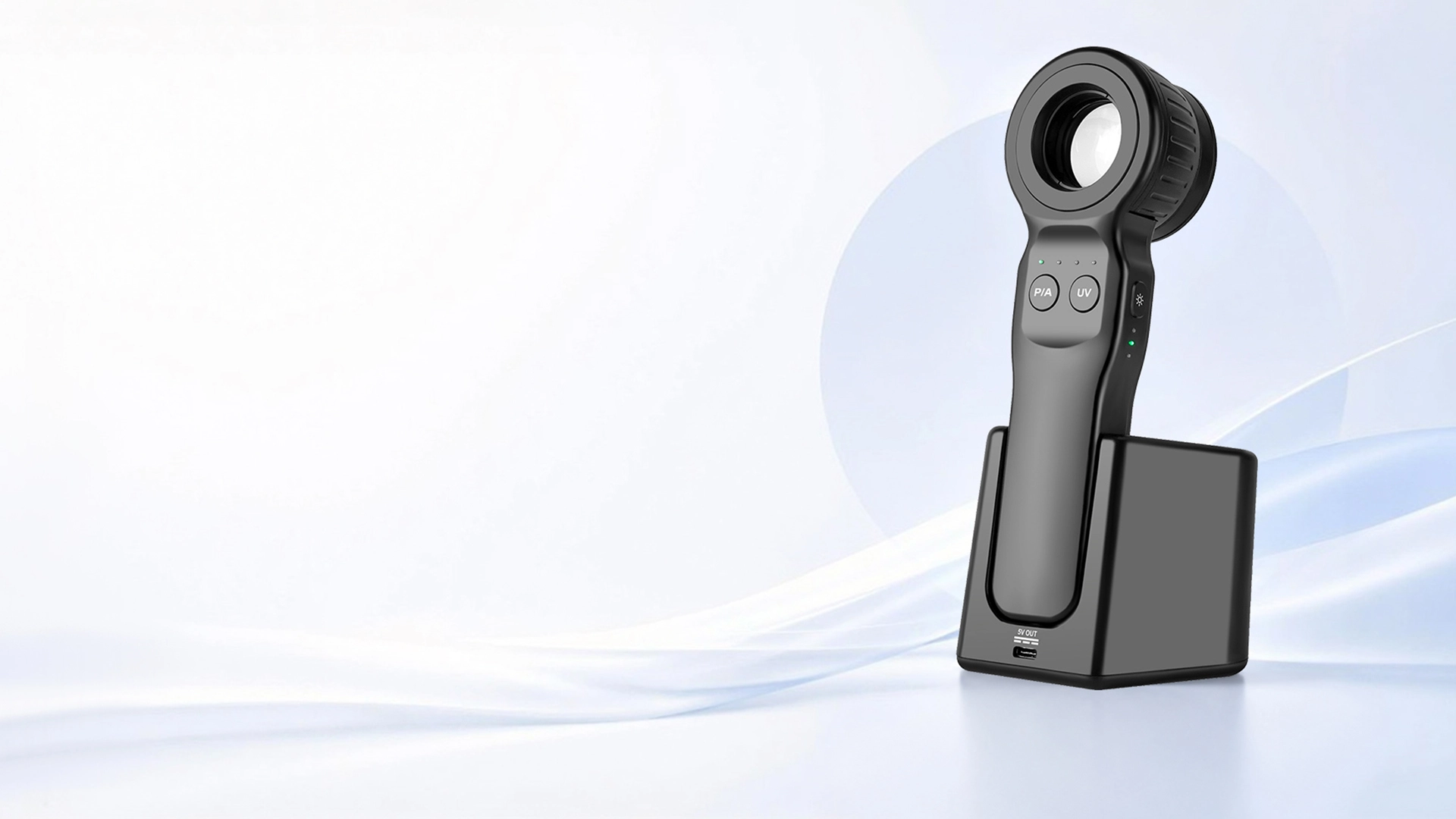
DE-5100 Advanced DermatoscopeSee More. Detect Earlier.
IBOOLO dermatoscopes combine advanced optical technology with versatile illumination and ergonomic design to deliver clear, detailed skin visualization for professional dermatology examination.


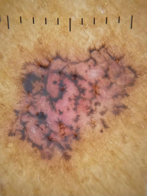
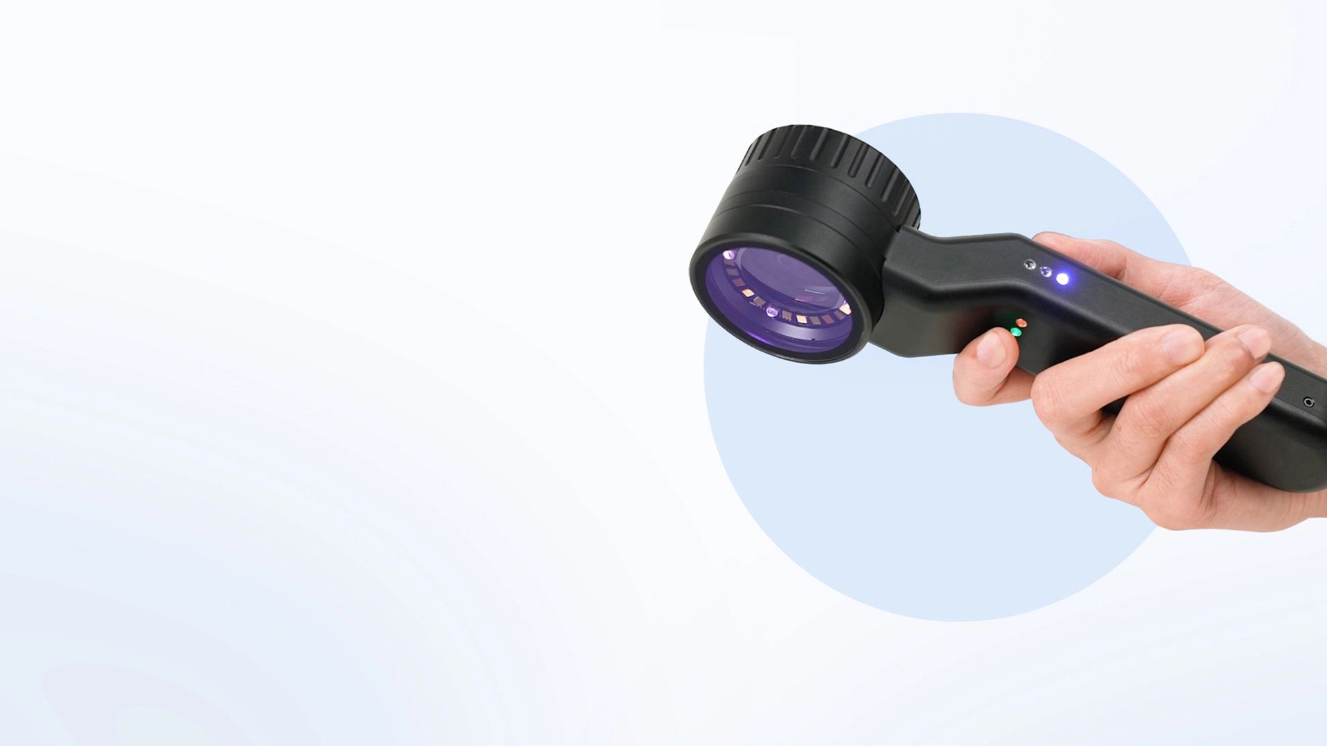
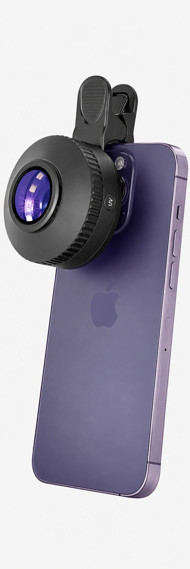
DE-4100 PRO
DE-4100 PRO Detachable DermatoscopeCapture More. Examine Better.
Provides a convenient, protable, and efficient approach for high-resolution, in-depth analysis of skin lesions, delivering clear visualization for confident clinical examination.

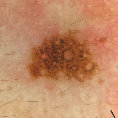
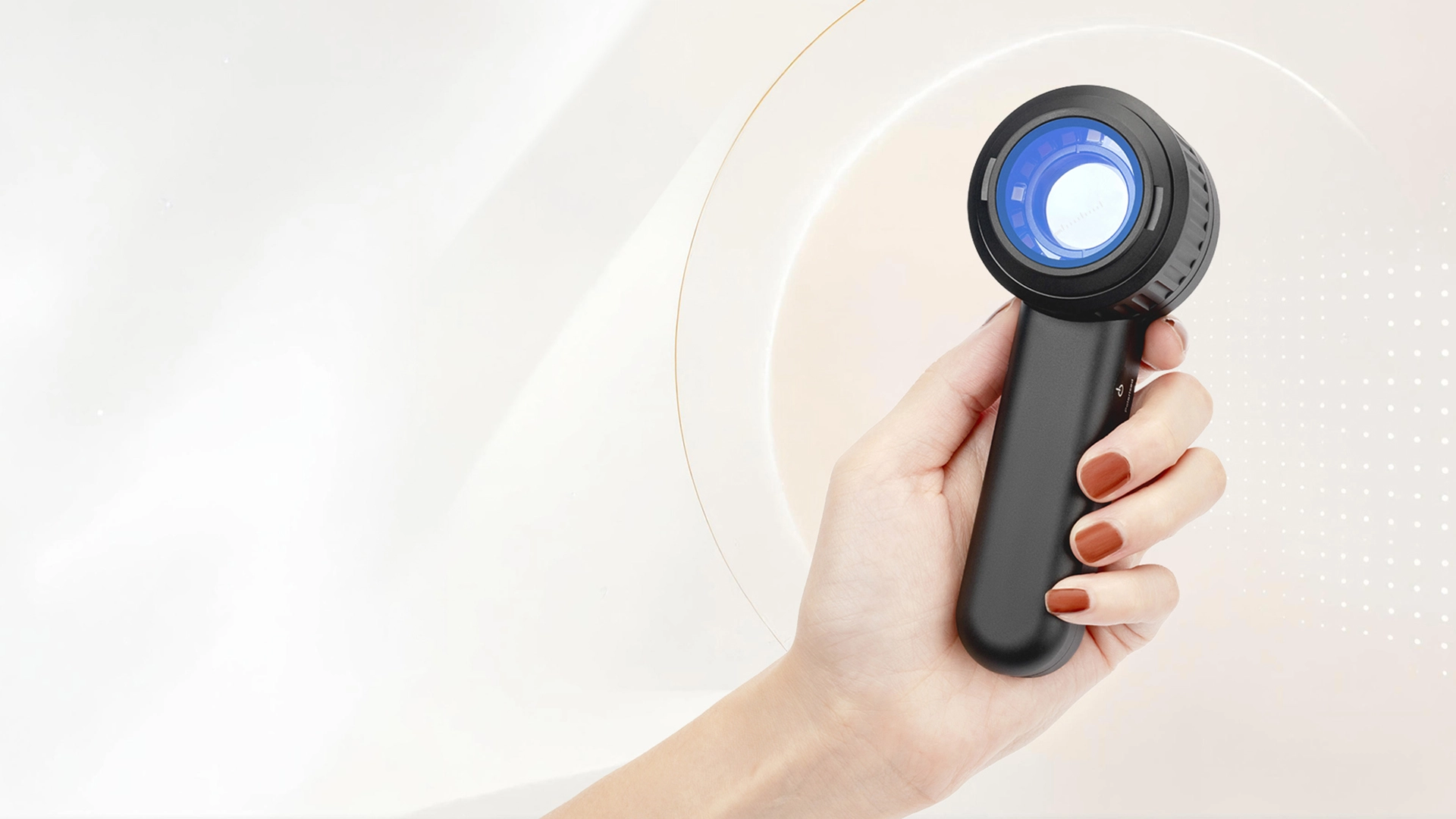
DE-3100 PRO
DE-3100 PRO Handheld DermatoscopeCapture More. Document Better.
Enables a more accurate, clear, and detailed examinition of the colors, shapes, and textures of skin lesions, combining portable design with high-resolution imaging for convenient digital dermoscopy and clinical documentation.


Explore IBOOLO Our Product Categories

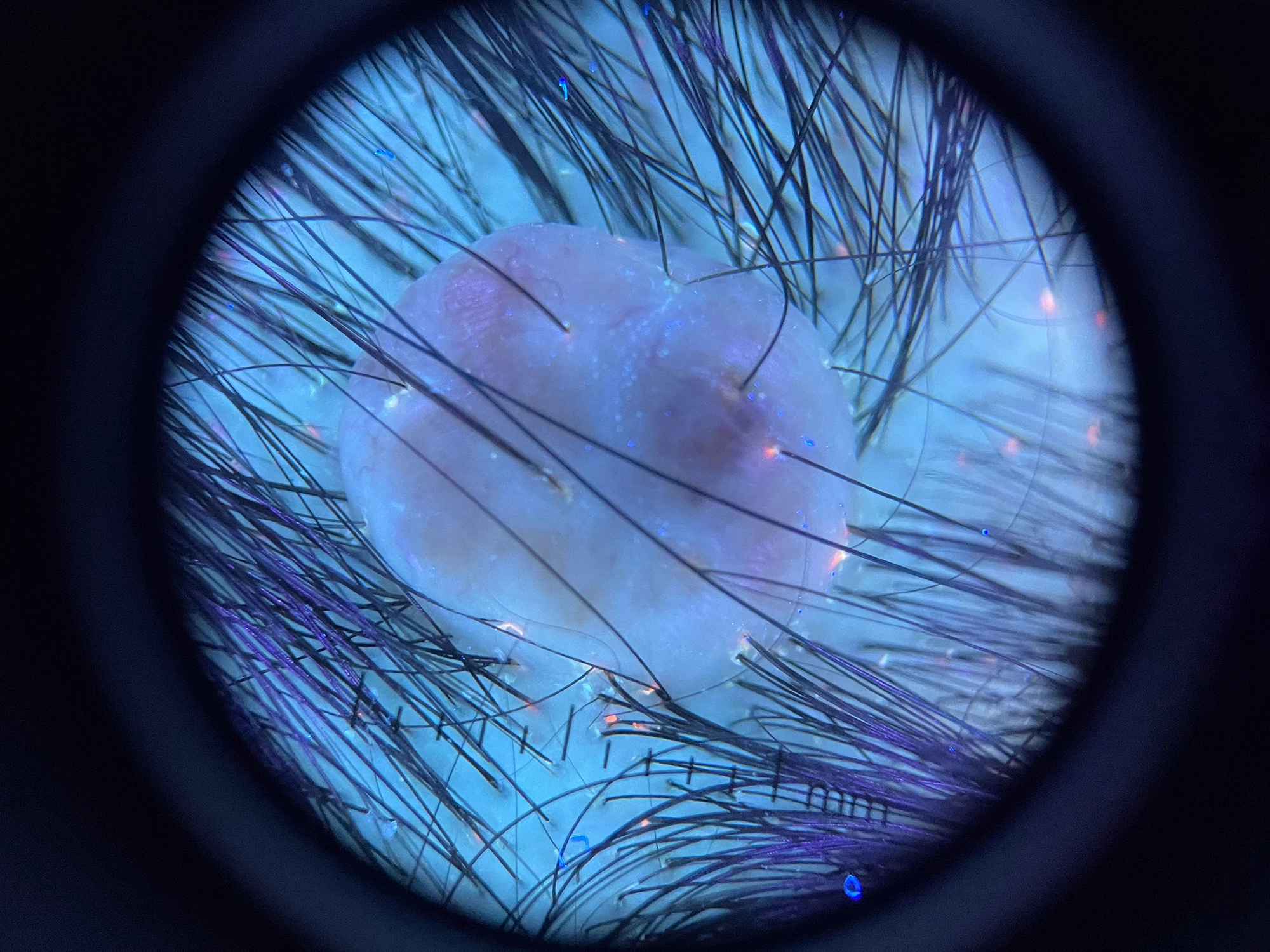
Dermatoscope

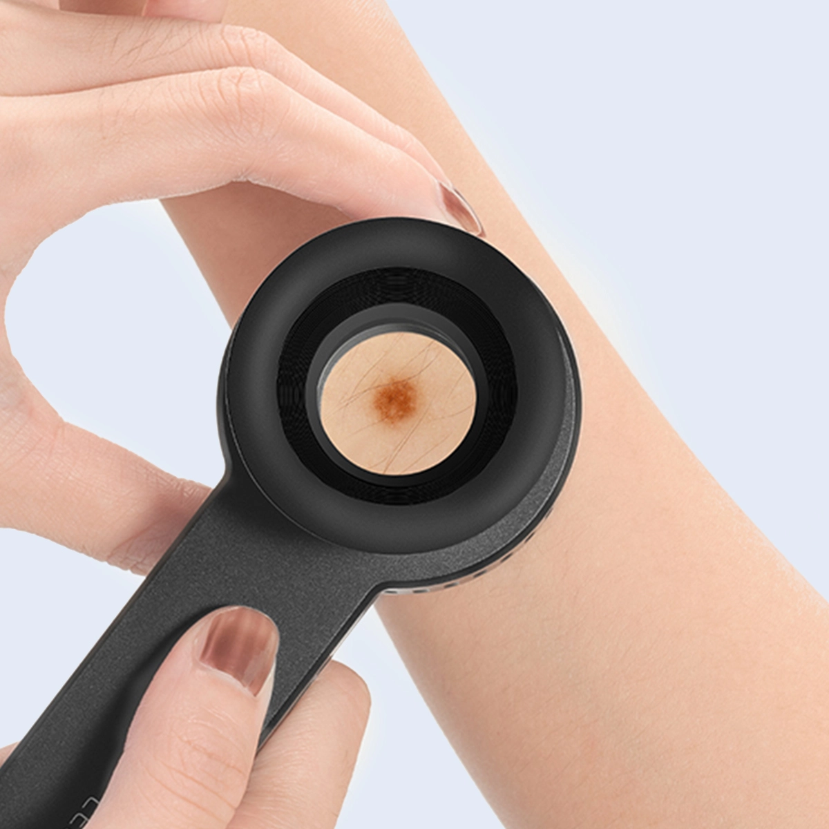
Woods Lamp

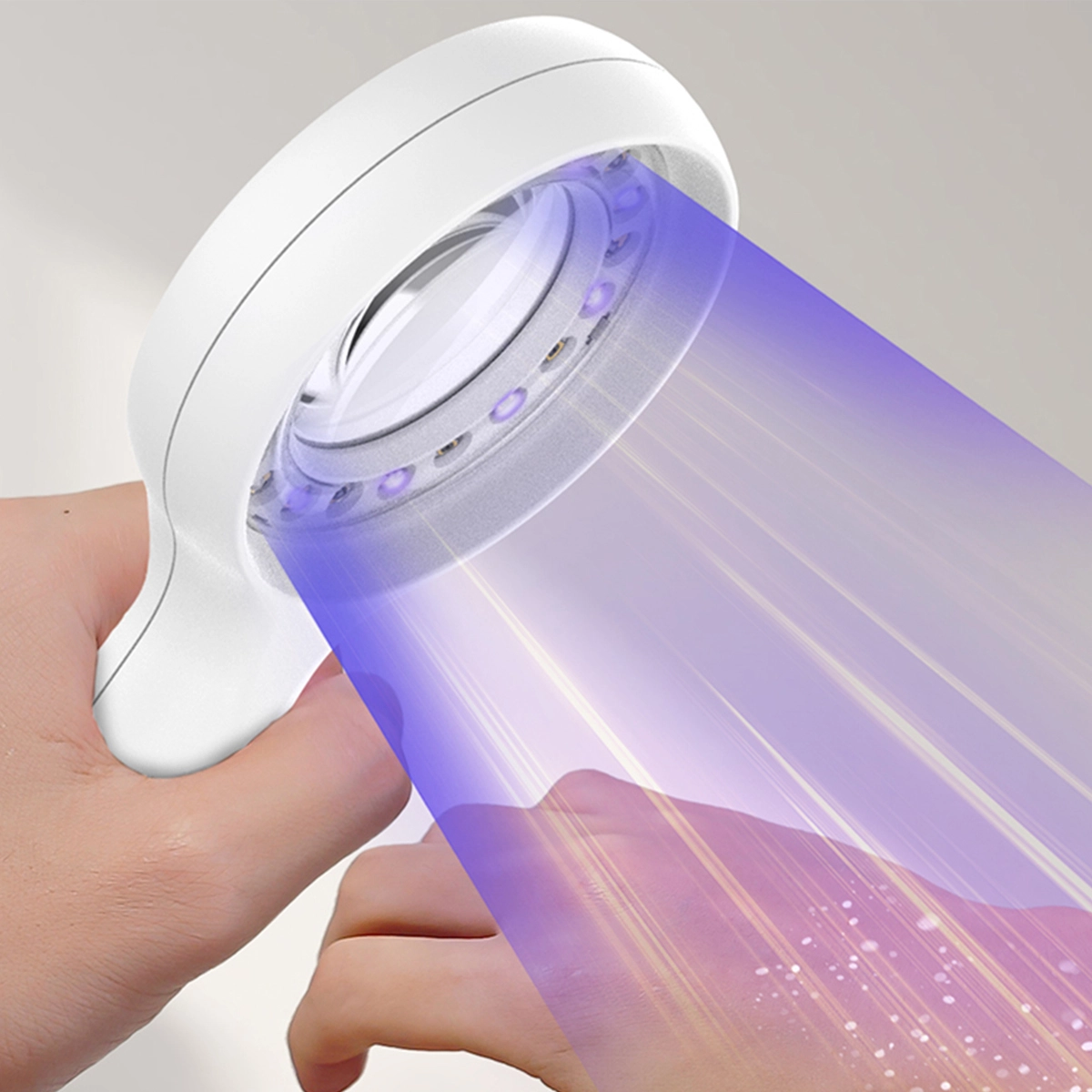
Phone & Camera Adapters
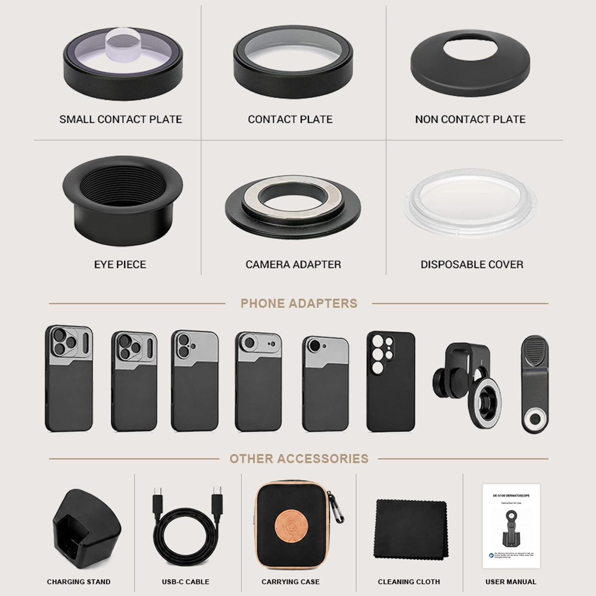

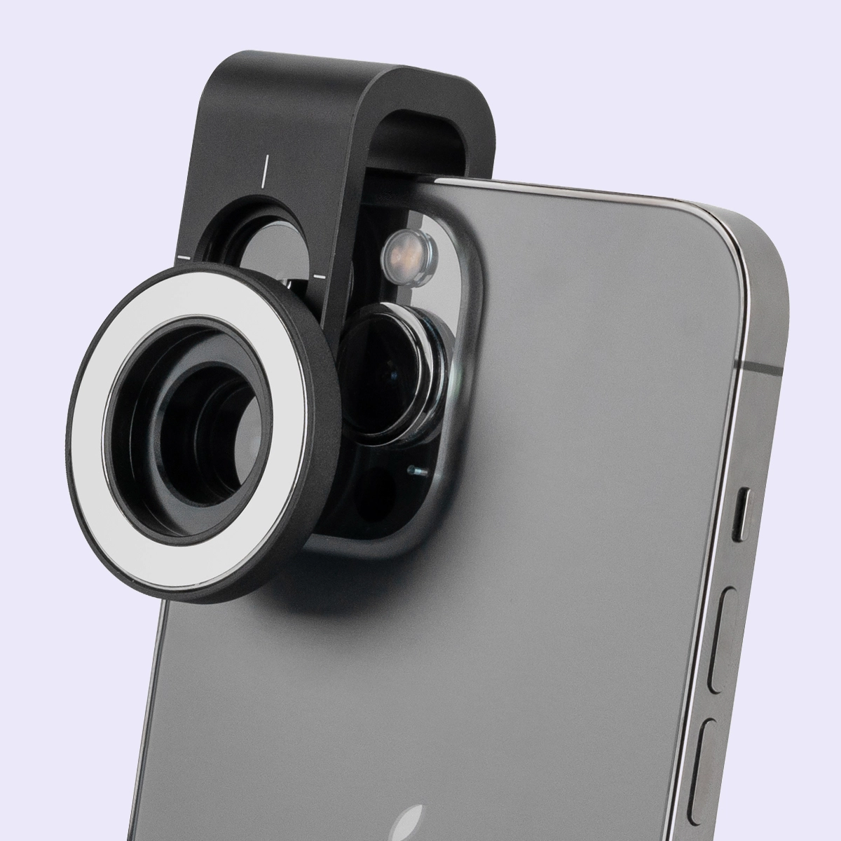
Accessories
Our Range IBOOLO Dermatoscope Series
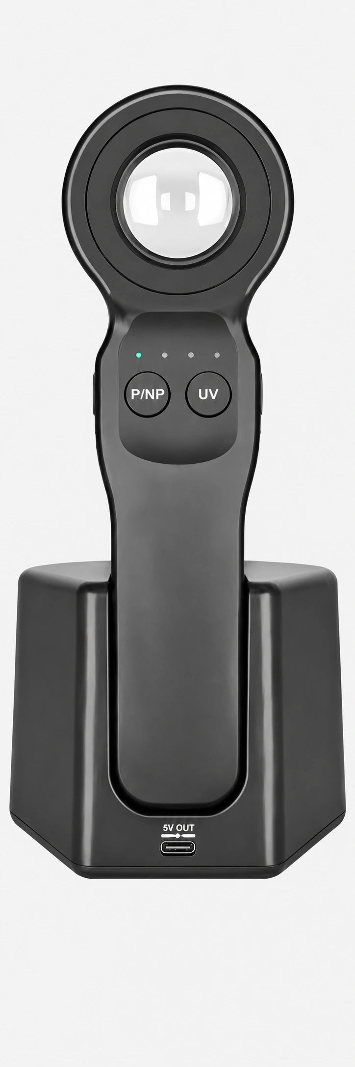
DE-5100 Dermatoscope
- 12x Magnification
- Multi-Polarization
- UV Illumination
- Wireless Charging
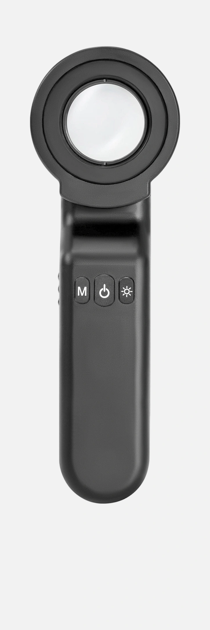
DE-4100 PRO Dermatoscope
- 32mm wide field of view
- 10 x Magnification
- Polarized Imaging
- Long Battery Life
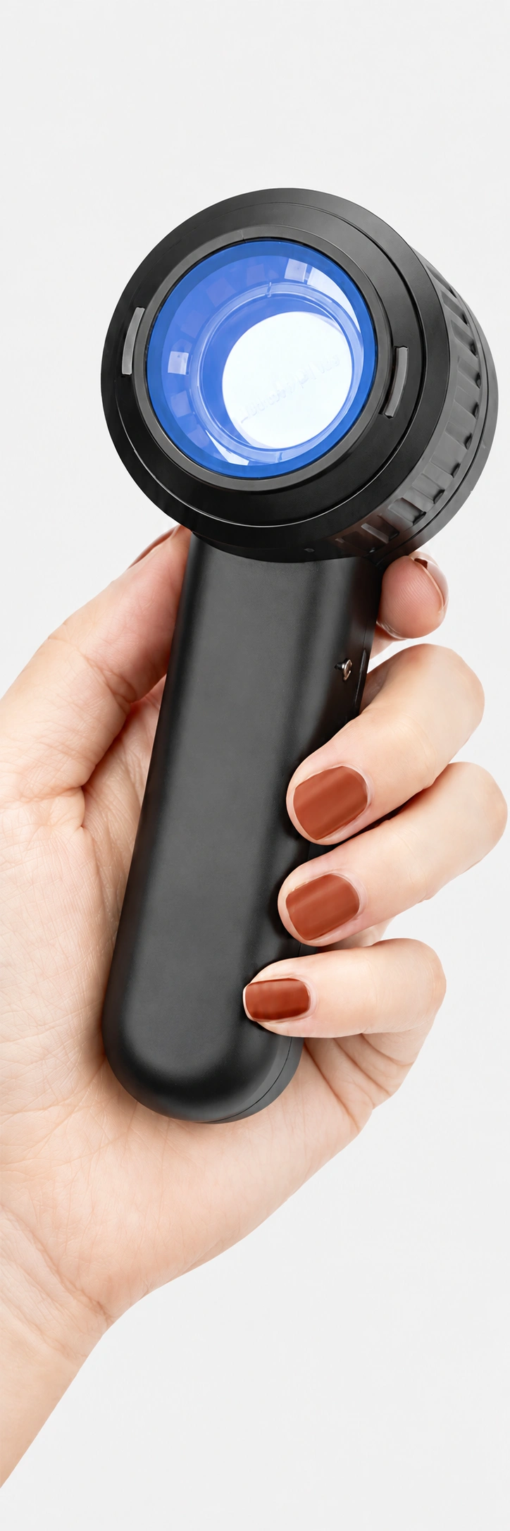
DE-3100 PRO Dermatoscope
- Smartphone Integration
- Portable Design
- UV Illumination
Compare Dermatoscopes Find the right dermatoscope for your practice
| Model | Magnification | Polarization | UV | Visual Field | Charging | Battery | Best For | |
|---|---|---|---|---|---|---|---|---|
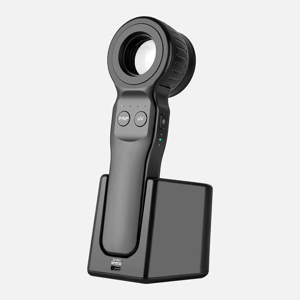 | DE-5100 | 12X | Cross + Parallel Polarized | 365nm | 32mm | Wireless | 1500mAh | Advanced Dermatology |
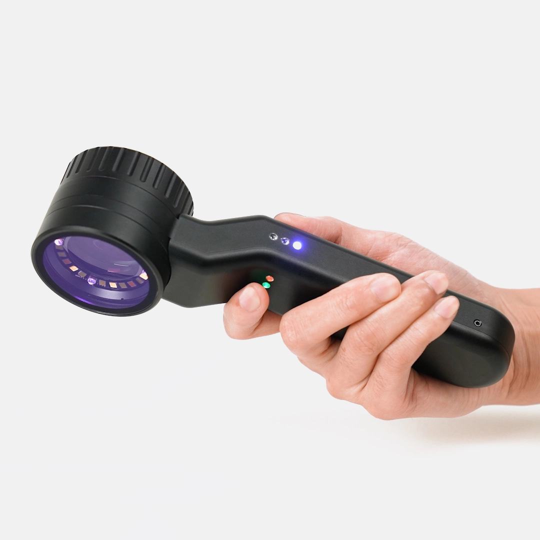 | DE-4100 PRO | 10X | Cross Polarized | 365nm | 32mm | Cable | 1500mAh | Professional Diagnostics |
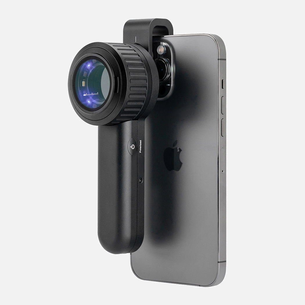 | DE-3100 PRO | 10X | Cross Polarized | 365nm | 25mm | Cable | 1000mAh | Gerneral Clinic Use |
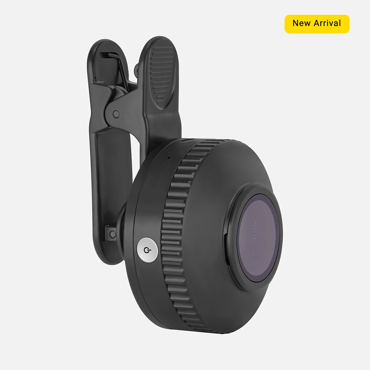 | DE-500 | 10X | Cross Polarized | 365nm | NA | Cable | 300mAh | Education, Training |
Our Latest Articles Find the latest dermatology news
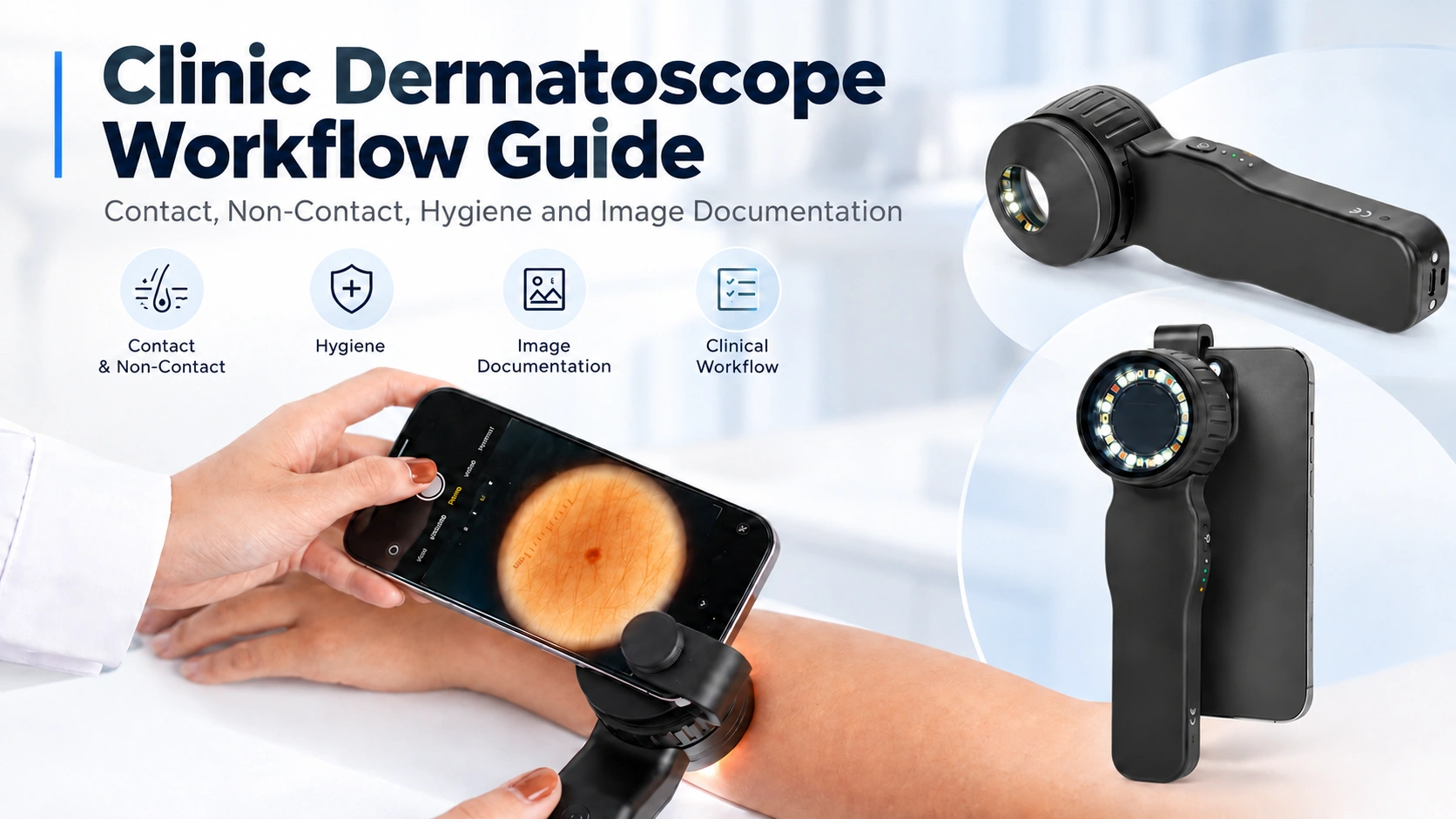
Clinic Dermatoscope Workflow Guide Contact, Non-Contact, Hygiene and Image Documentation
A dermatoscope may be a compact handheld instrument, but using it efficiently in a clinic involves more than switching on the light and examining a lesion. A practical clinic workflow dermatoscope setup needs to consider how the device is prepared, whether contact or non-contact observation is appropriate, how different illumination…
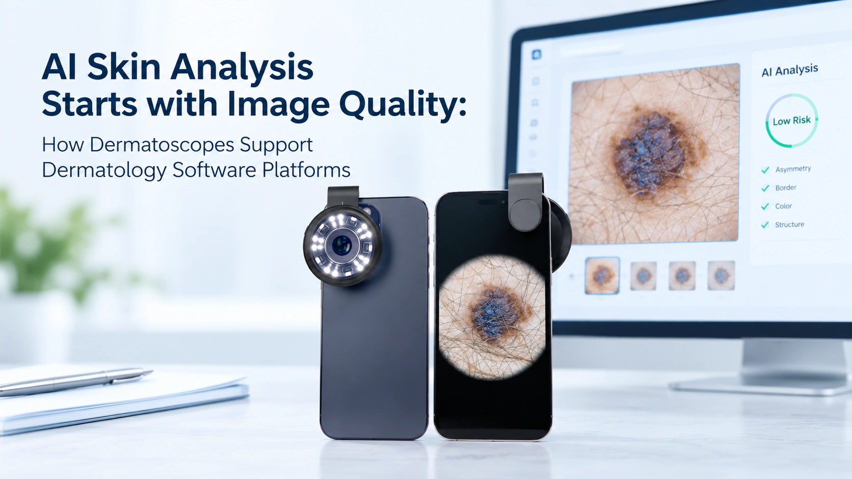
AI Skin Analysis Starts with Image Quality: How Dermatoscopes Support Dermatology Software Platforms?
At IBOOLO, we see AI skin analysis as a workflow that begins before an algorithm processes an image. Dermatology software can classify, compare or analyze only the visual information contained in the image it receives. If the photograph is out of focus, affected by glare, captured from an inconsistent distance…
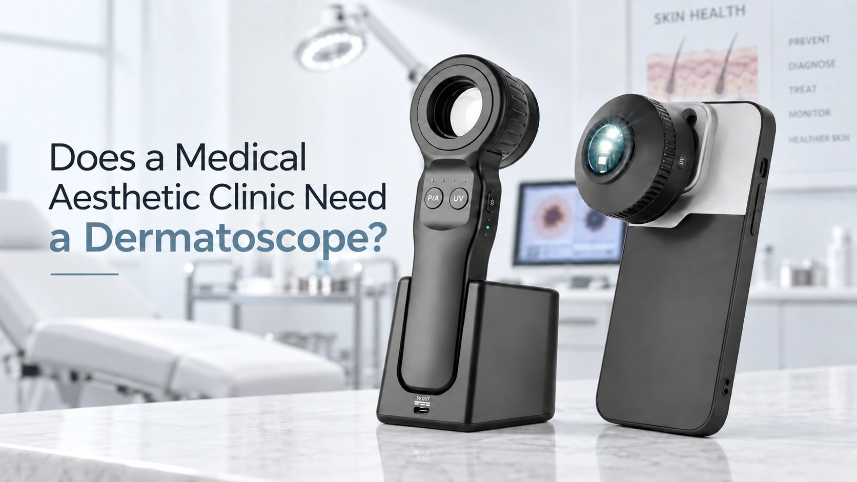
Does a Medical Aesthetic Clinic Need a Dermatoscope?
Medical aesthetic clinics work directly with the skin every day. During a consultation for laser treatment, hair removal, pigmentation management or another cosmetic procedure, a client may suddenly point to a mole or dark spot and ask whether it is normal, whether it has changed, or whether it could indicate…
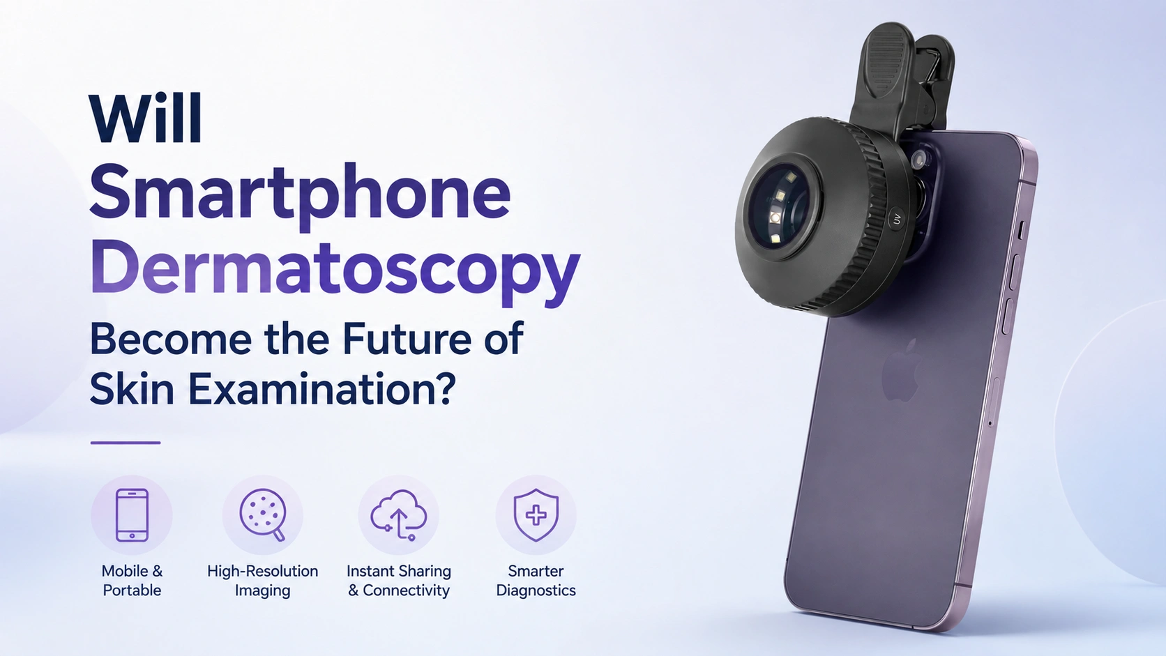
Will Smartphone Dermatoscopy Become the Future of Skin Examination?
Smartphones are now widely used to capture, store, share, and review medical images. In dermatology, this has created growing interest in smartphone dermatoscopy. By combining a dermatoscope with a smartphone camera, clinicians can capture magnified images of skin lesions without relying only on visual memory or written descriptions. However, the…
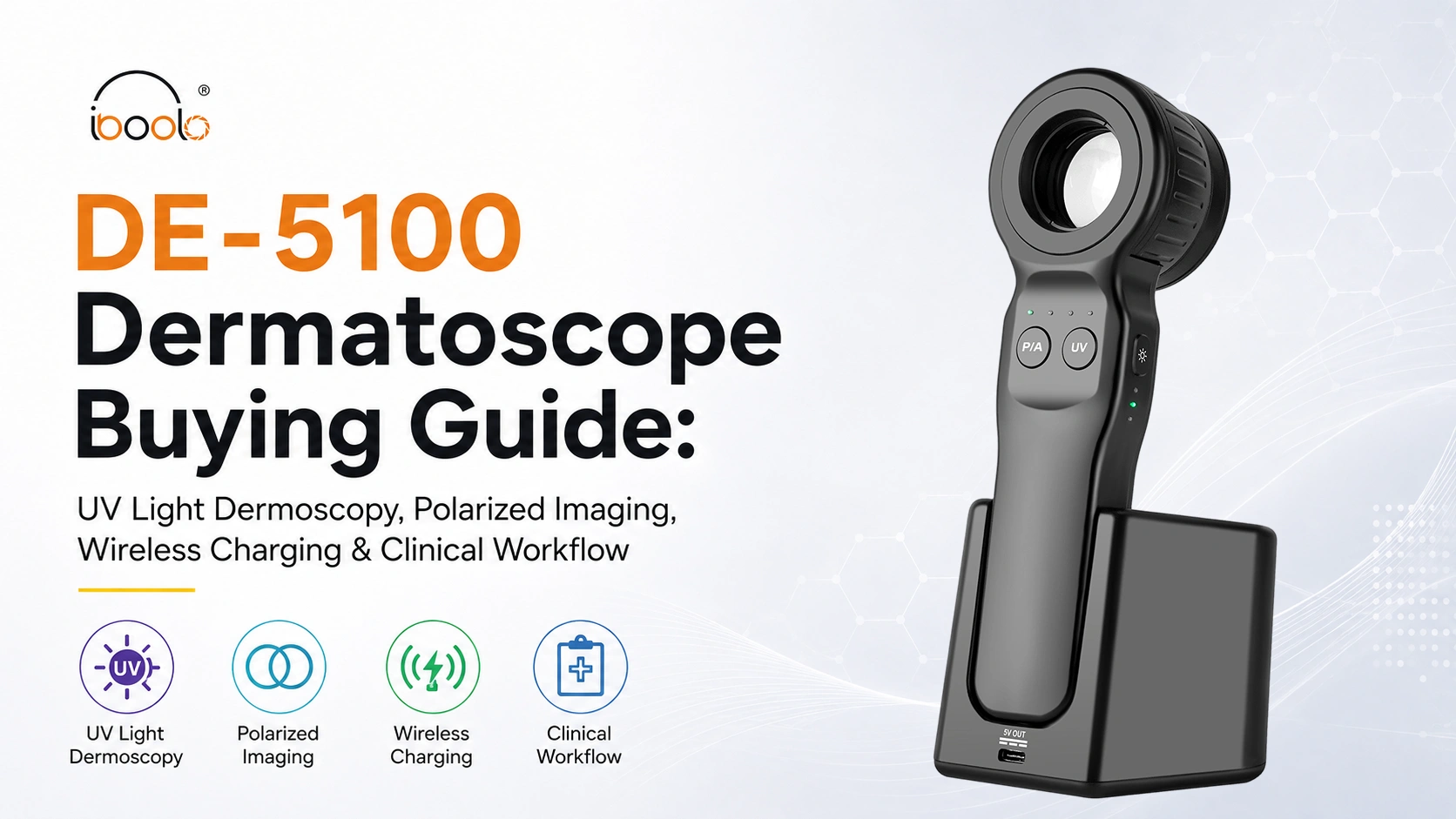
DE-5100 Dermatoscope Buying Guide: UV Light Dermoscopy, Polarized Imaging, Wireless Charging & Clinical Workflow
As dermoscopy continues to evolve, clinicians are looking for more than magnification alone. Modern dermatological practice increasingly emphasizes efficient clinical workflows, standardized image documentation, and the ability to observe skin structures under different illumination modes. Whether in dermatology clinics, primary care settings, educational institutions, or aesthetic practices, healthcare professionals often…
Clinic Dermatoscope Workflow Guide Contact, Non-Contact, Hygiene and Image Documentation
A dermatoscope may be a compact handheld instrument, but using it efficiently in a clinic involves more than switching on…
AI Skin Analysis Starts with Image Quality: How Dermatoscopes Support Dermatology Software Platforms?
At IBOOLO, we see AI skin analysis as a workflow that begins before an algorithm processes an image. Dermatology software…
Does a Medical Aesthetic Clinic Need a Dermatoscope?
Medical aesthetic clinics work directly with the skin every day. During a consultation for laser treatment, hair removal, pigmentation management…
Will Smartphone Dermatoscopy Become the Future of Skin Examination?
Smartphones are now widely used to capture, store, share, and review medical images. In dermatology, this has created growing interest…
DE-5100 Dermatoscope Buying Guide: UV Light Dermoscopy, Polarized Imaging, Wireless Charging & Clinical Workflow
As dermoscopy continues to evolve, clinicians are looking for more than magnification alone. Modern dermatological practice increasingly emphasizes efficient clinical…
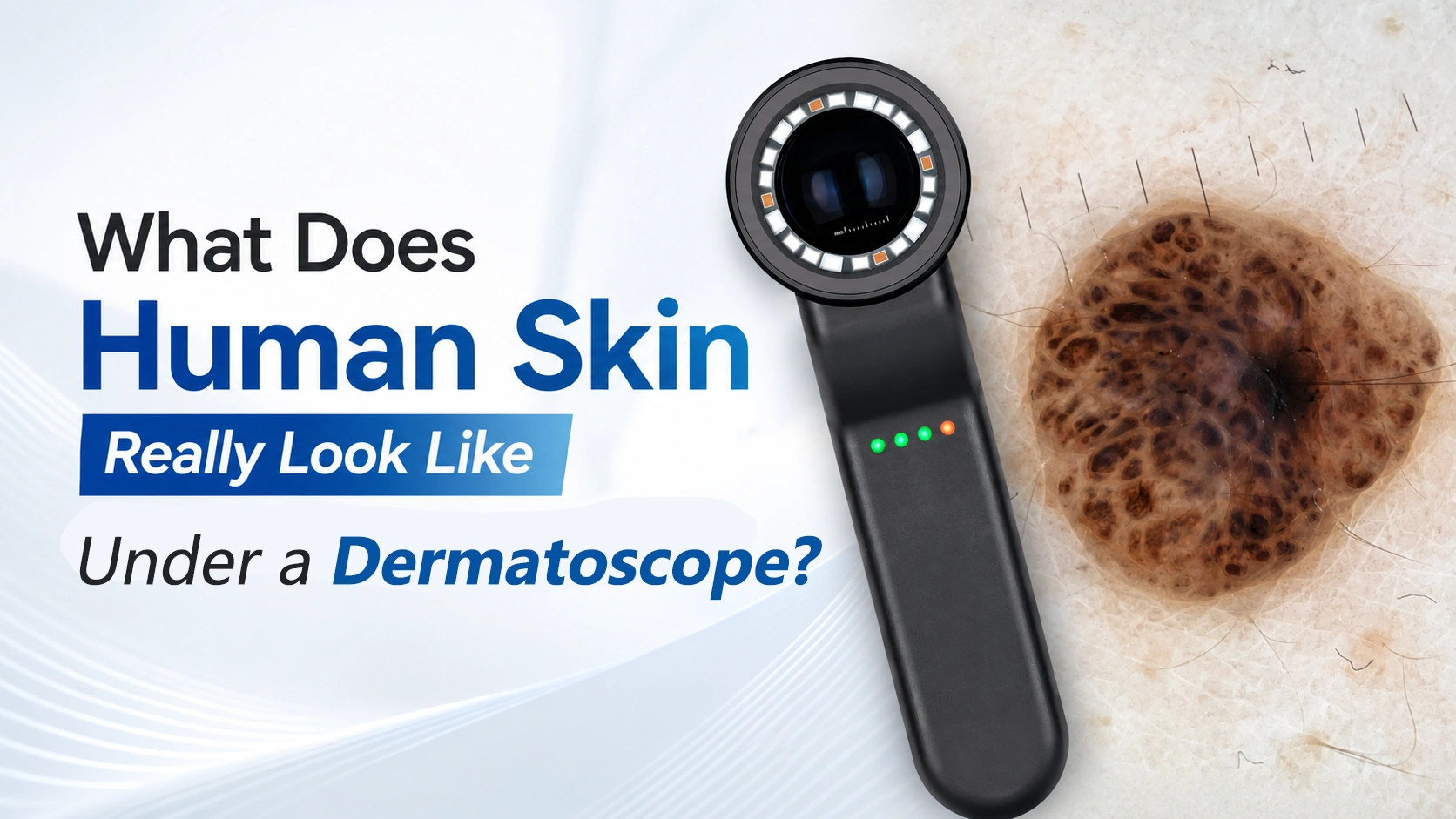
What Does Human Skin Really Look Like Under a Dermatoscope?
Most people judge a skin lesion with the naked eye. We notice its color, size, shape, or whether it has…
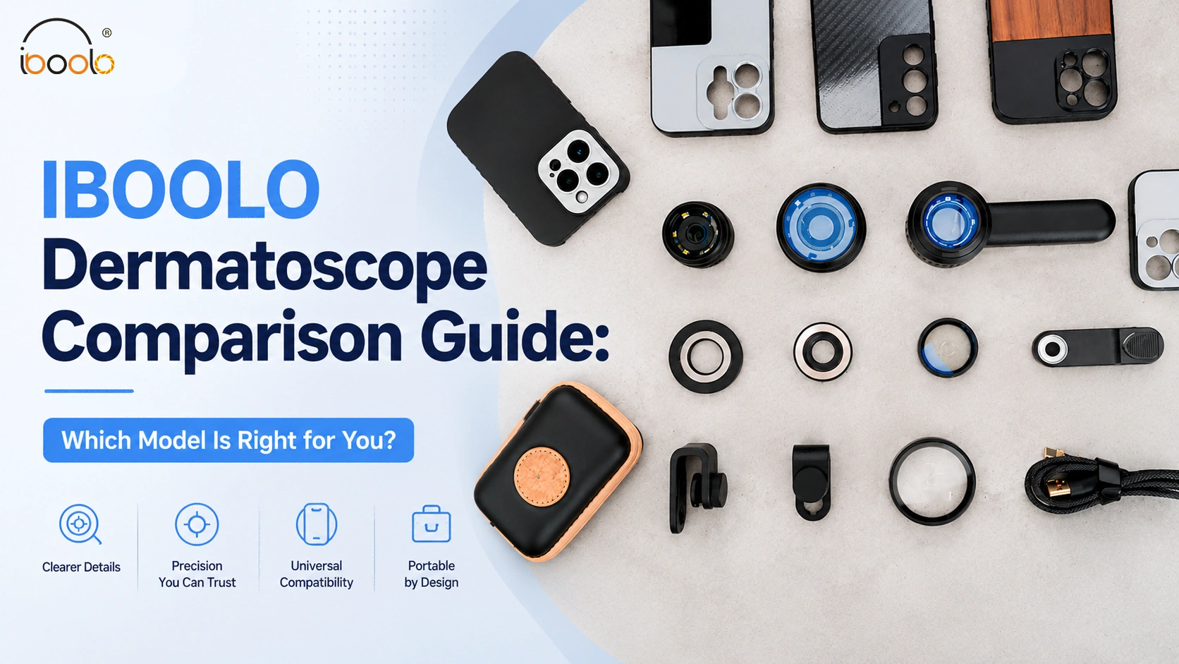
IBOOLO Dermatoscope Comparison Guide: Which Model Is Right for You?
Dermatoscopes have become important tools for skin observation and image documentation. Different users have different needs. Some prefer direct visual…
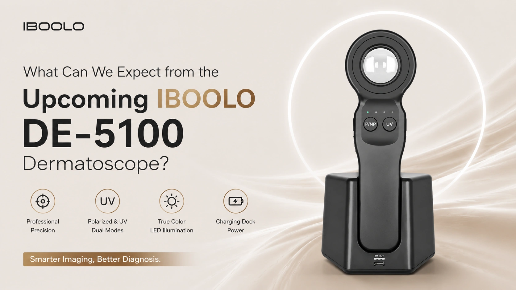
What Can We Expect from the Upcoming IBOOLO DE-5100 Dermatoscope?
Dermoscopy has become an important tool in modern dermatology. Numerous studies published in international medical journals have shown that dermoscopy…
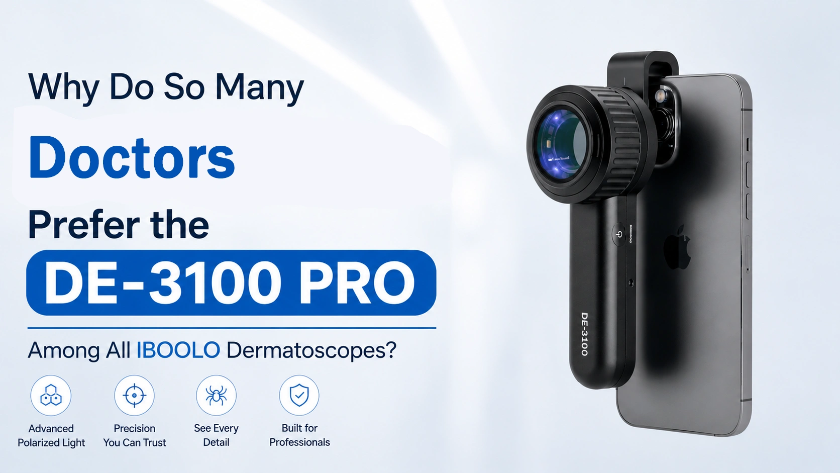
Why Do So Many Doctors Prefer the DE-3100 PRO Among All IBOOLO Dermatoscopes?
Dermatoscopy has transformed the clinical evaluation of skin lesions, providing a non-invasive bridge between macroscopic examination and histopathological analysis. Within…
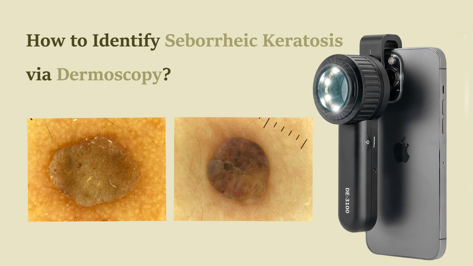
The Most Common Benign Lesion: How to Identify Seborrheic Keratosis via Dermoscopy?
Seborrheic keratosis (SK) represents one of the most frequent reasons for dermatological consultations globally. Often referred to as senile warts…
Why Choose IBOOLO Trusted by Professionals, Chosen Worldwide
IBOOLO develops and manufactures high-quality dermatoscopes with advanced optics, reliable performance, and professional support for healthcare professionals worldwide.

Advanced Optics
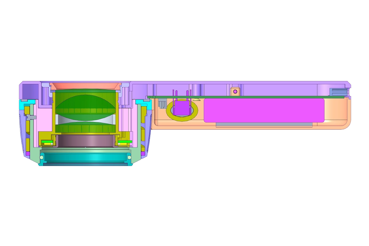
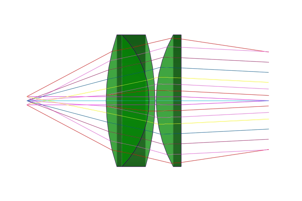
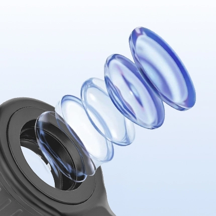
High-resolution all-glass optics with anti-reflection coating deliver excep-tional clarity and accurate visualiza-tion of skin structures.

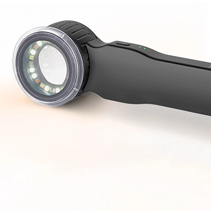
Multiple Illumination Modes
Cross, parallel, amber polarization, and 365nm UV illumination provide versatile imaging options for different clinical applications.

Cross Polarized

Parallel Polarized

Amber Polarized

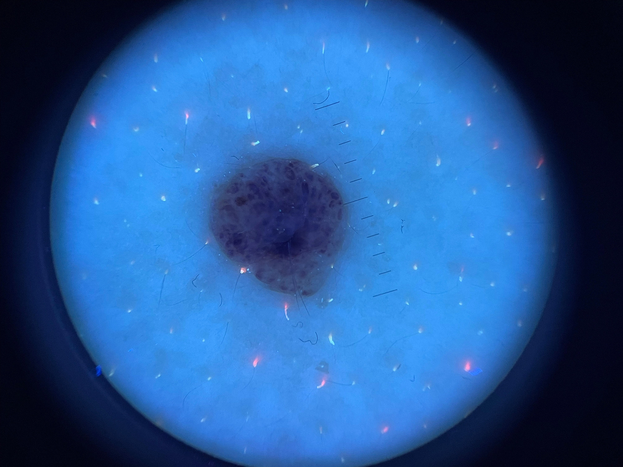
365nm UV Light

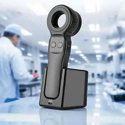
ISO 13485 Manufacturing
Manufactured under and ISO 13485 certified quality management system to ensure consistent quality, reliability and regulatory compliance.

Global Support
Dedicated technical support and after-sales service to assist distributors, clinics, and healthcare professionals worldwide.


Reliable Quality
Built to last. Backed by rigor.

Innovative Technology
Designed for better diagnosis.

Trusted by Professionals
Preferred by dermatologists worldwide.

Worldwide Delivery
Fast and secure shipping to 100+ countries.
Clinical Applications Built for Every Skin Assessment
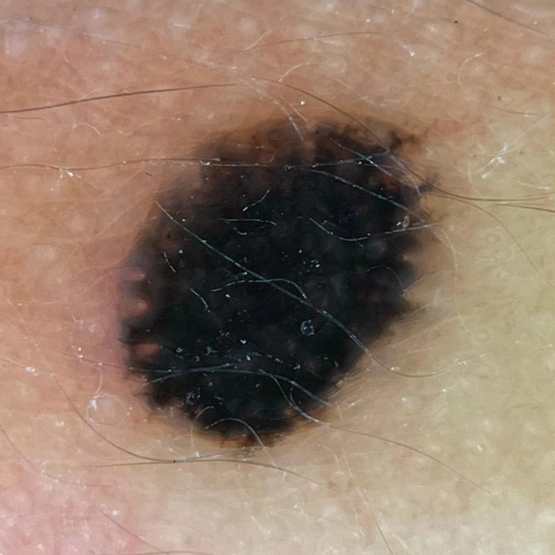
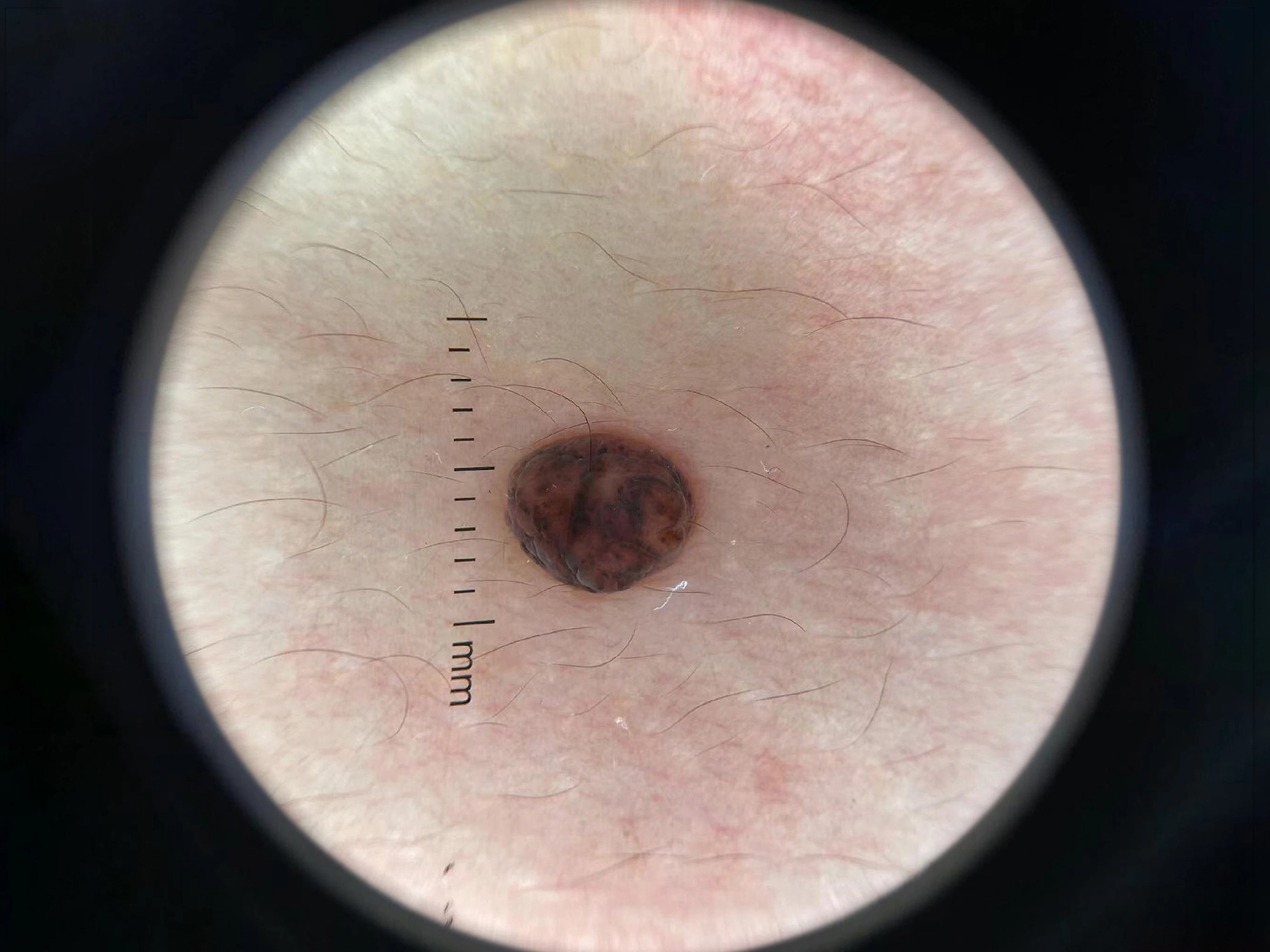
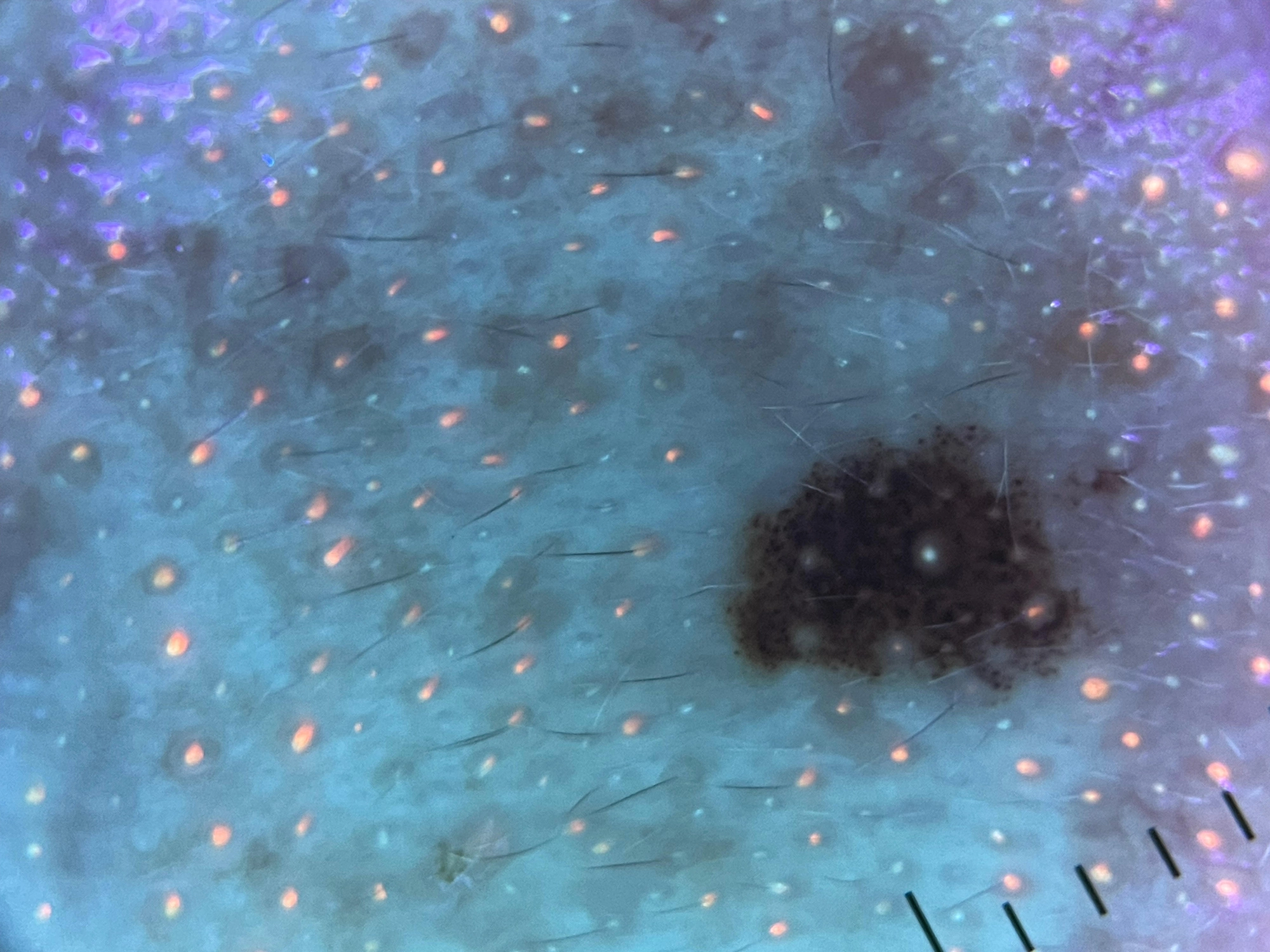
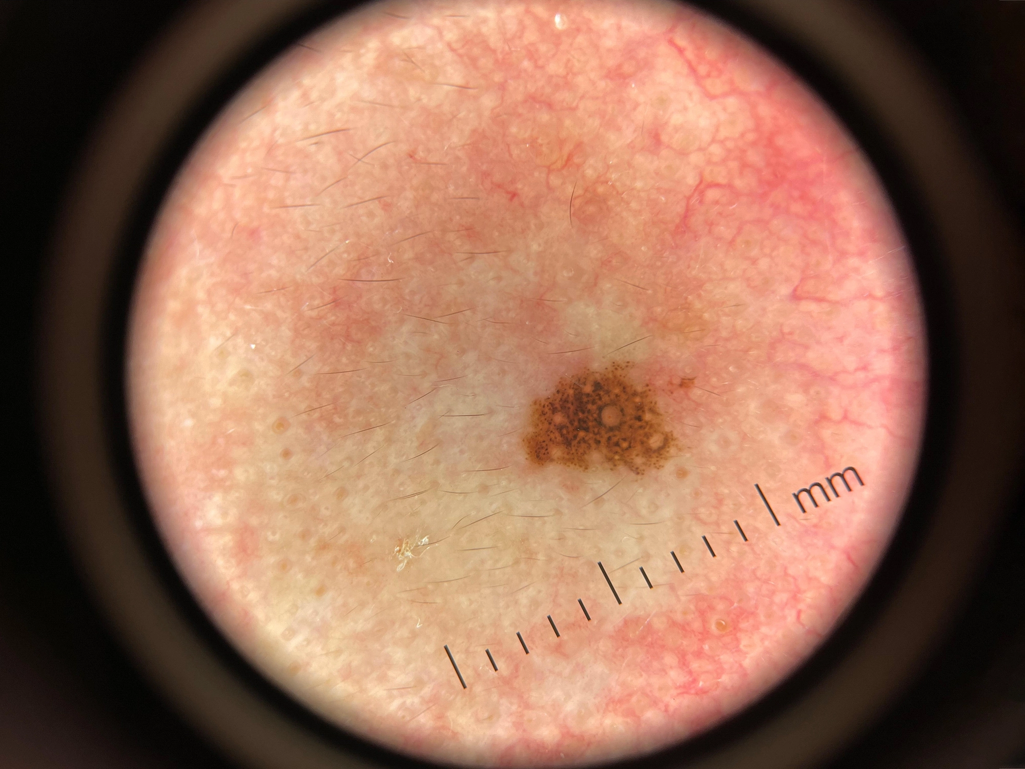
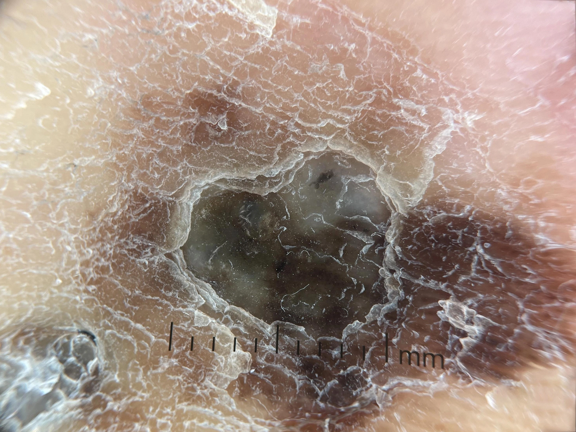
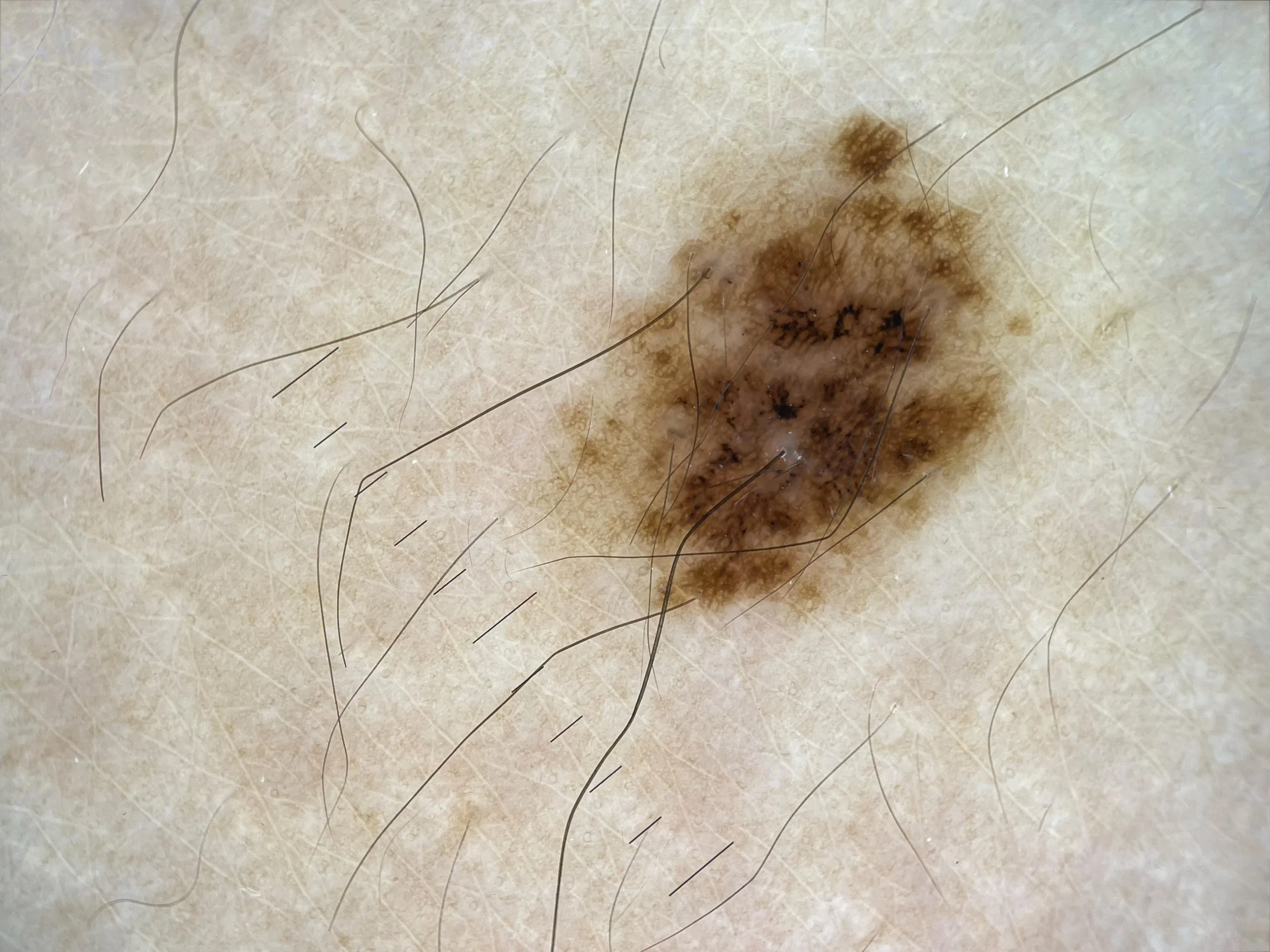
Pigmented Lesions
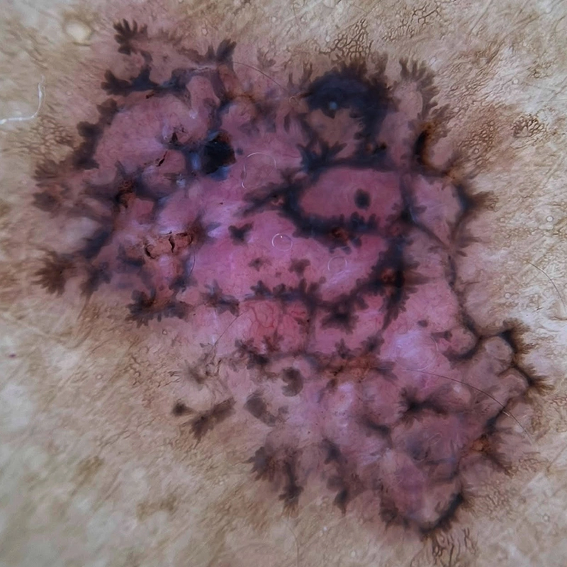
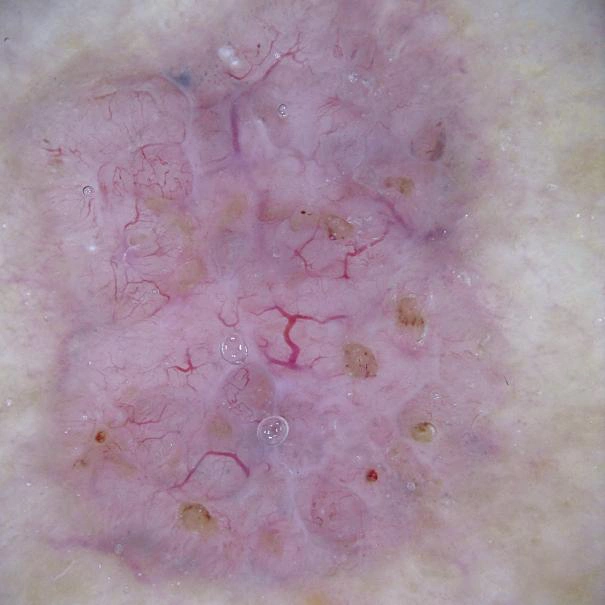
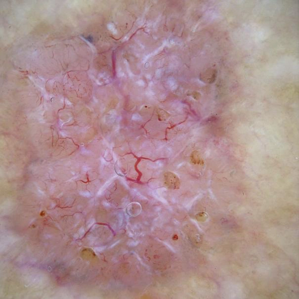
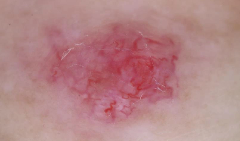
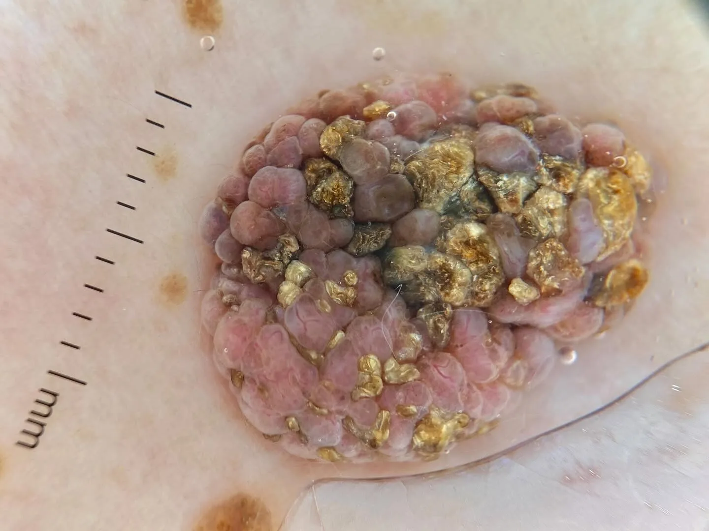
Basal Cell Carcinoma (BCC)
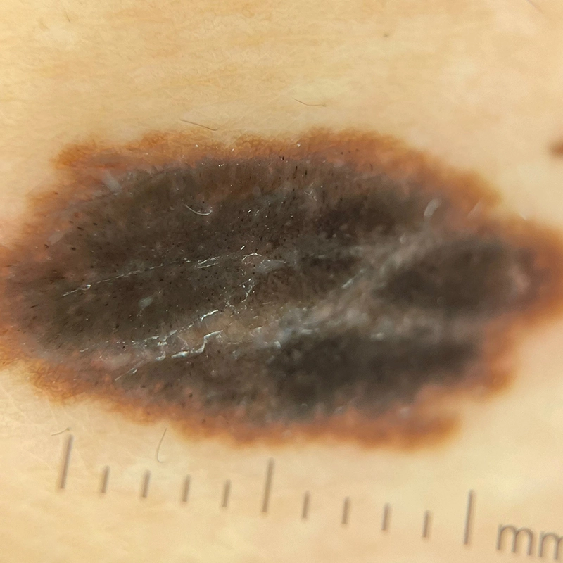
Suspicious Pigmented Lesions
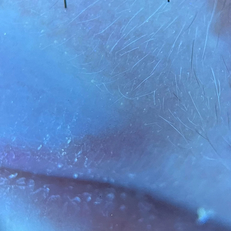
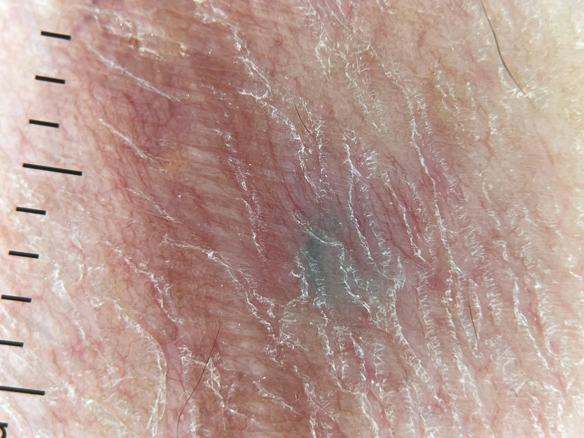
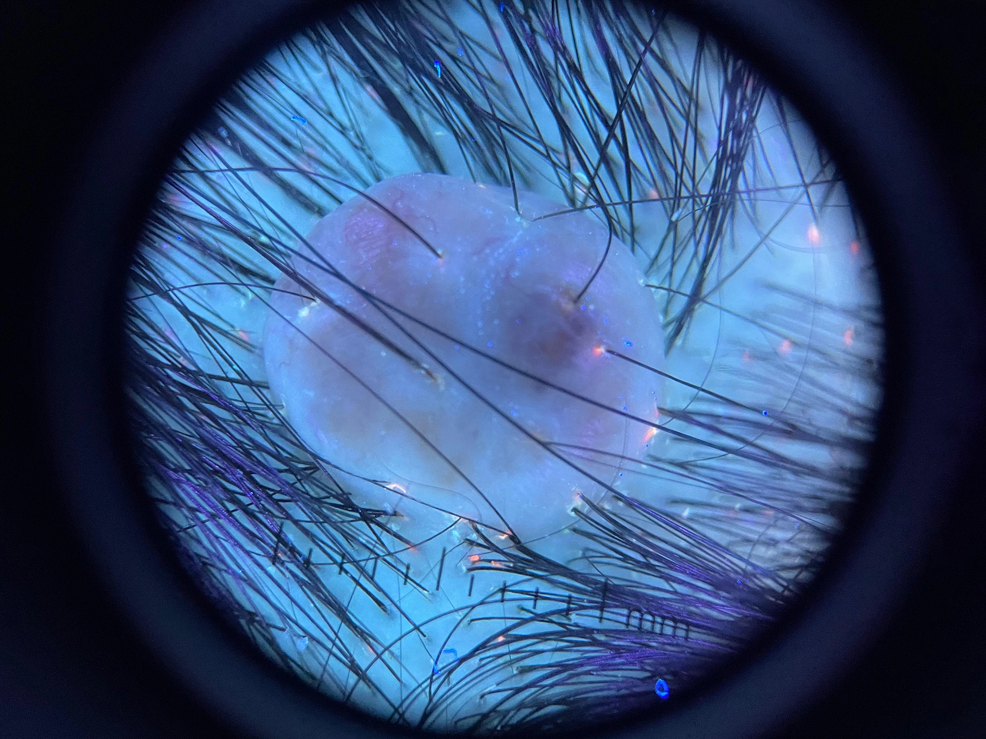
Vitiligo Assessment
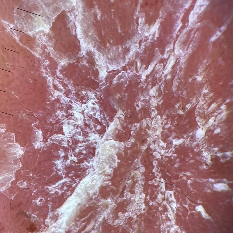
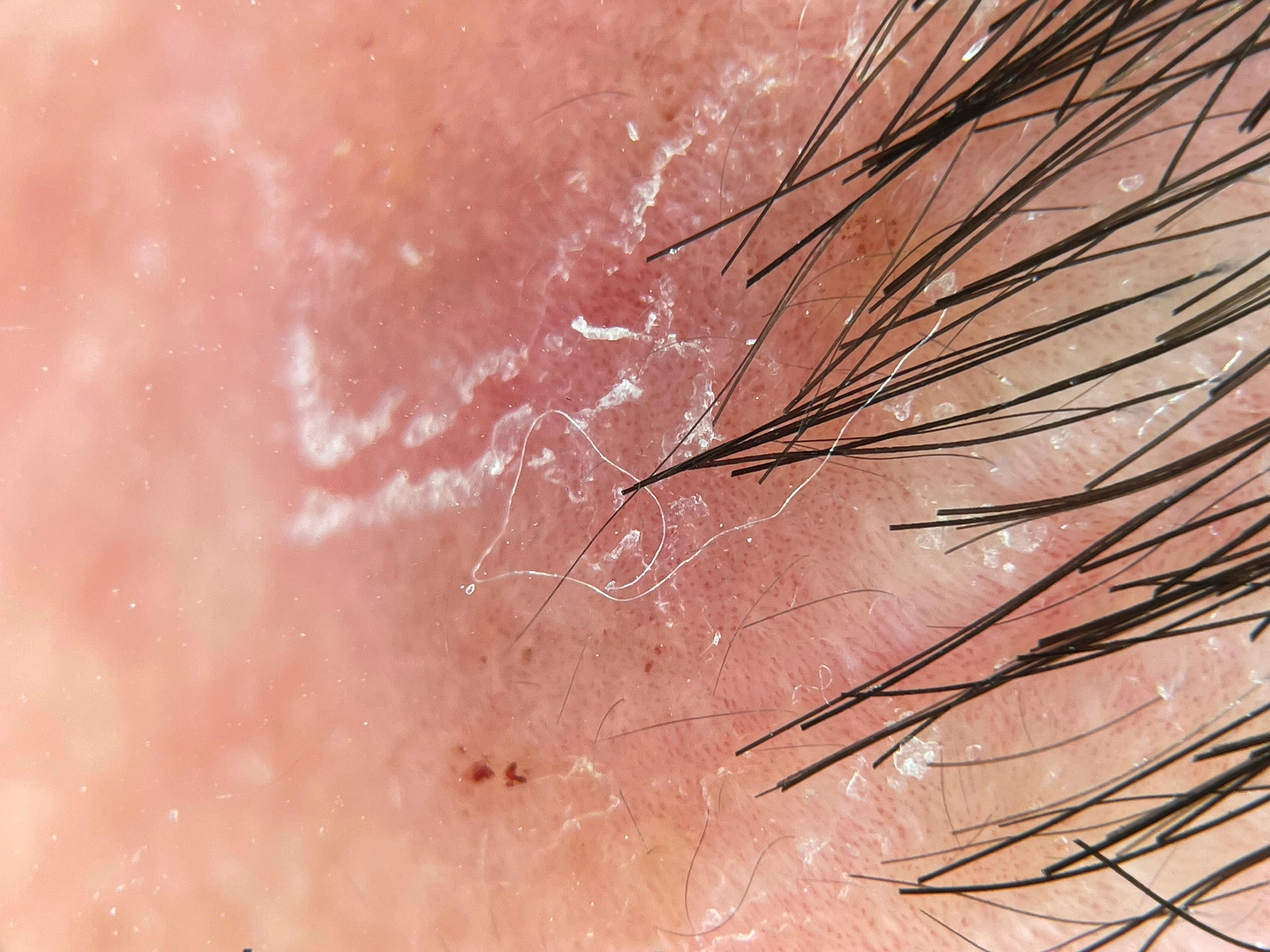
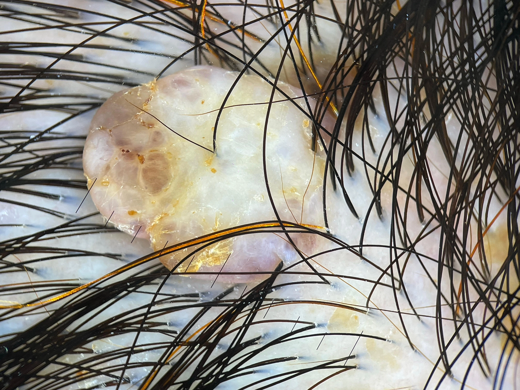
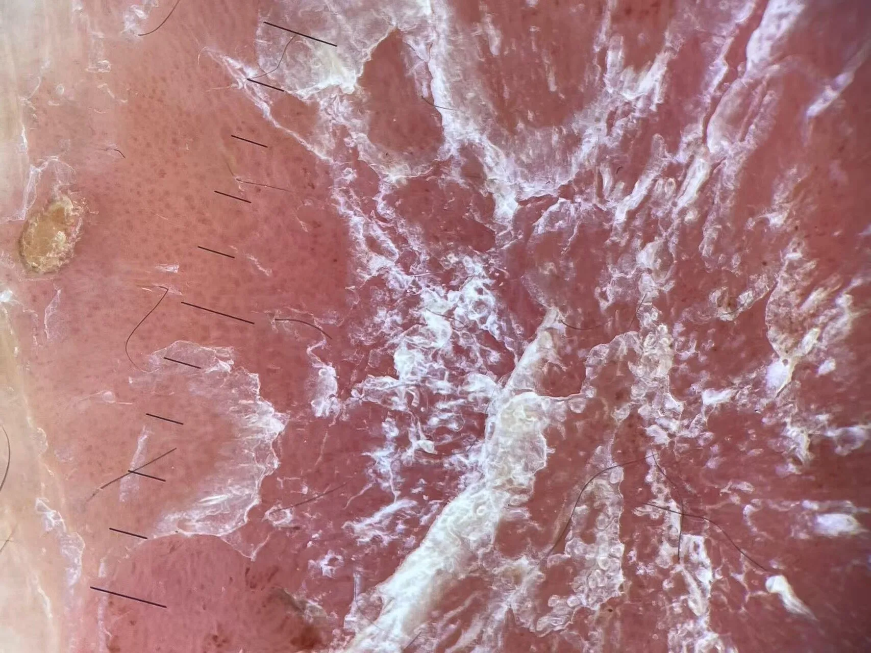
Psoriasis Assessment
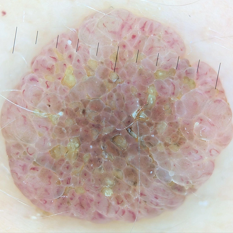
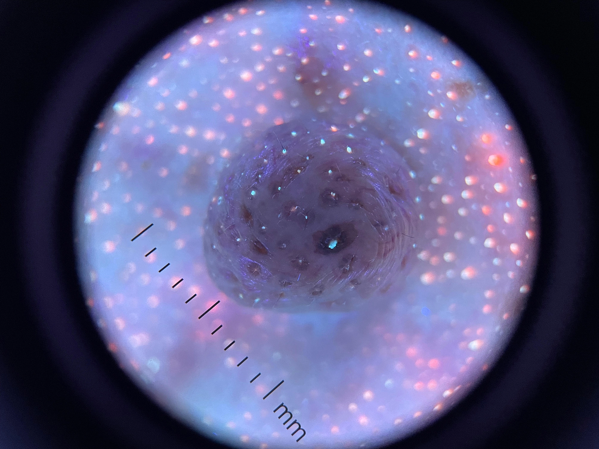
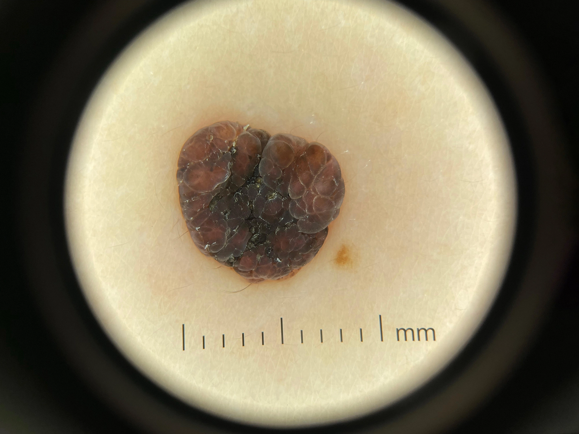
Benign Nevus Examination

Accepted Payments










100% Satisfaction Guaranteed
We offer
"7-day No Questions Asked Return or Exchange" and "2-year Product Warranty" to every product purchased.
Contact Us or Learn More.