


DE-400 Dermatoscope – IBOOLO
Premium Digital Dermatoscope Manufacturer China - IBOOLO
People May Ask
melanoma. Although most melanomas are brown or black in color, some might have pink, tan, or even white hues. Not all melanomas are spherical like regular moles; some have colored sections. They could proliferate swiftly or even penetrate the skin around them.
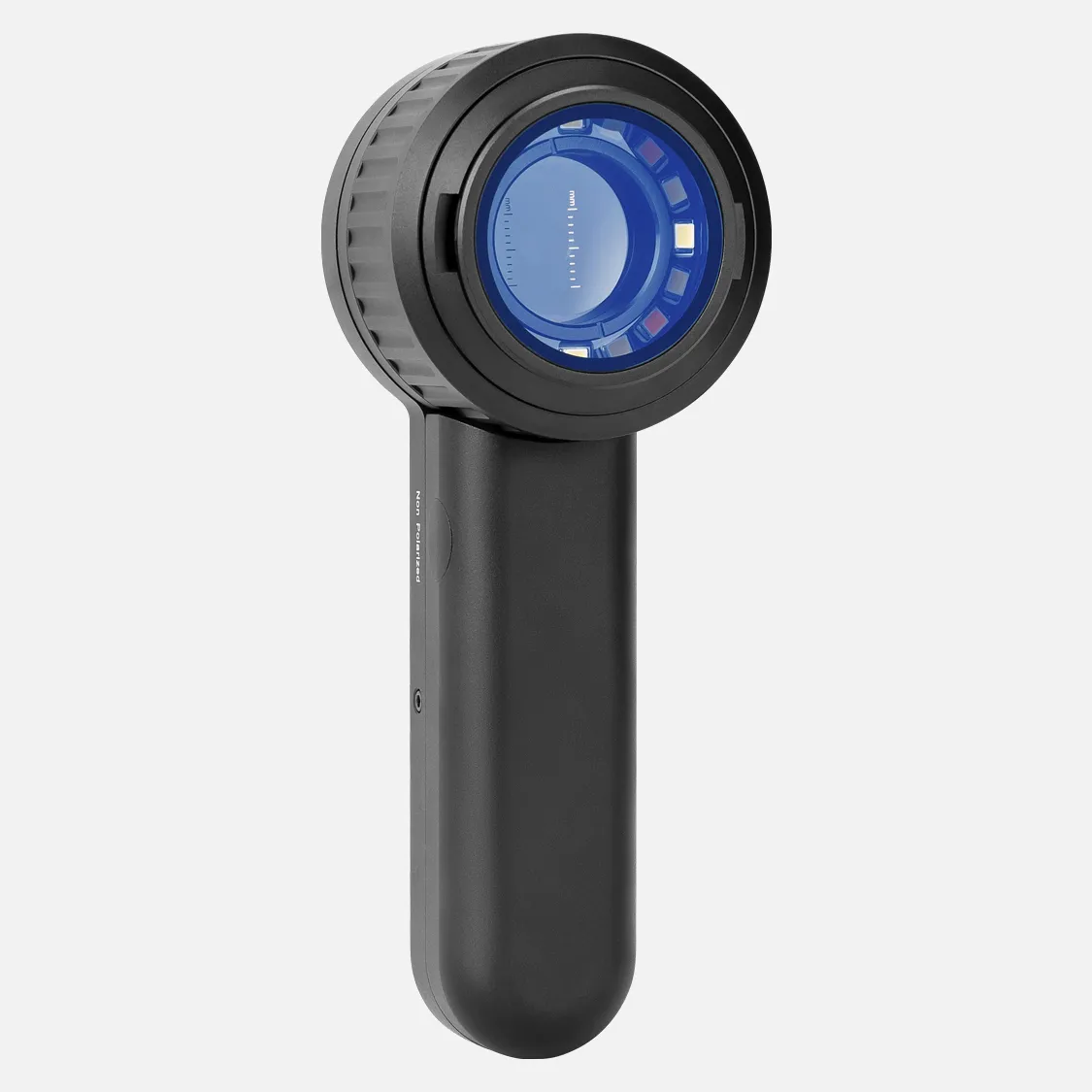
With a dermatoscope, features can be evaluated down to the reticular dermis, and pictures can be taken for further analysis. Transillumination of a lesion for high magnification examination and visualization of minor details is the fundamental idea behind dermoscopy.
As they expand, melanomas may begin as flat patches. 4. If you can feel it, you should get it checked out, even if some moles can also be raised. Occasionally, when assessing melanoma, the "E" in the ABCDE guidance refers to "evolving." This is a result of the gradual changes in melanomas' size, shape, and color.
Thick pigmented lines around appendageal apertures, commonly referred to as rhomboidal structures, are the hallmarks of lentigo maligna's dermoscopic features.Unevenly colored follicular apertures.Instead,globules and dots of slate grey.
Is melanoma possible from a common mole? Melanoma, the most dangerous kind of skin cancer, is extremely uncommon to develop from a common mole. Those with numerous little or multiple large moles are more likely to acquire melanoma, even though common moles are not malignant (1).
Not all moles with scabs are malignant. But sometimes, scabby moles are malignant. If you are unable to link the scabbing to a recognized skin injury, it is crucial that you have them examined.
Initial clinical symptoms and misdiagnoses. In our study, the initial physician visit resulted in an erroneous diagnosis for 30% of the melanomas. This aligns with the outcomes of previous cohorts. Fortin and colleagues discovered a 25% rate of initial misdiagnosis, but Bristow and Acland reported a 33% rate of wrong diagnosis.
pigment that has spread into the surrounding skin from the spot's edge. Redness or a fresh enlargement outside the mole's boundaries. Modification in feeling, including pain, sensitivity, or itching. A mole's surface may change, becoming scaly, leaking, bleeding, or developing a bulge or bump.
An essential tool for melanoma early detection is melanoma mole mapping. Dermatologists may detect possible problems and offer prompt treatment by routinely checking your skin for changes.
With a dermatoscope, features can be evaluated down to the reticular dermis, and pictures can be taken for further analysis. Transillumination of a lesion for high magnification examination and visualization of minor details is the fundamental idea behind dermoscopy.
Digital Dermatoscope Products
Anykit Digital Otoscope with a gyroscope, 4.5-inch screen, 3.9mm ear scope camera with six lights, 32GB card, and an ear wax removal tool Allows for the capture of photos and videos

Dino-Lite AM3113T USB Digital Microscope with 0.3 MP, 10x - 50x, 230x Optical Magnification, Microtouch, Measurement, and Discontinued

SupereyesB005+1~200X Portable USB Digital Microscope, Endoscope Loupe, Otoscope Magnifier with 11mm Health Kit Tripod Diameter

Digital video otoscope/earscope Firefly DE550 Wireless

USB-C and Lightning Compatible Digital Microscope
The VMS-007-DX2 is a USB digital microscope with 5 Megapixels, x300 magnification, the ability to capture and record photos and videos, up to 2592 x 1944 resolution, 8 LEDs, and an adjustable stand.

The adult 4.3-inch IPS coin microscope with 1000X magnification is made by Hayve.Eight programmable LED coin collection suppliesCompatible with WindowsTF Card, 32GB
Digital Portable Microscope with 8 LED Magnifier, Manually Adjustable for Computer Phone and Tablet, 50X-500X 0.3MP USB Magnification

手持式 USB 数码显微敜带金属支枴,便携式高清 1000 倍放大检查摄像头,带 8 个 LED 灯,怂用于 Android Mac Windows 电脑
Vividia Digital Borescope Microscope with 12mm Diameter OTG Android and USB PC Compatible with a Professional Multi-Functional Metal Stand
Hot Products


News & Blog


Top Reviews
Edmond D.
Fantastic watch, excellent numbers are visible at night, and Italian is a language that is easy to imitate. I really like this watch.
Rhisian
The lampshade really highlights the nursery motif. It's really gorgeous and precisely what we were looking for—the green is a little bit lighter than it appears in the photo, more like a light sage.
Mrs K Frampton
Though they appear smaller in the photo, these are quite adorable and a really affordable way to alter the appearance of a regular lightbulb. To add a special touch, I painted a couple of these as well???????


