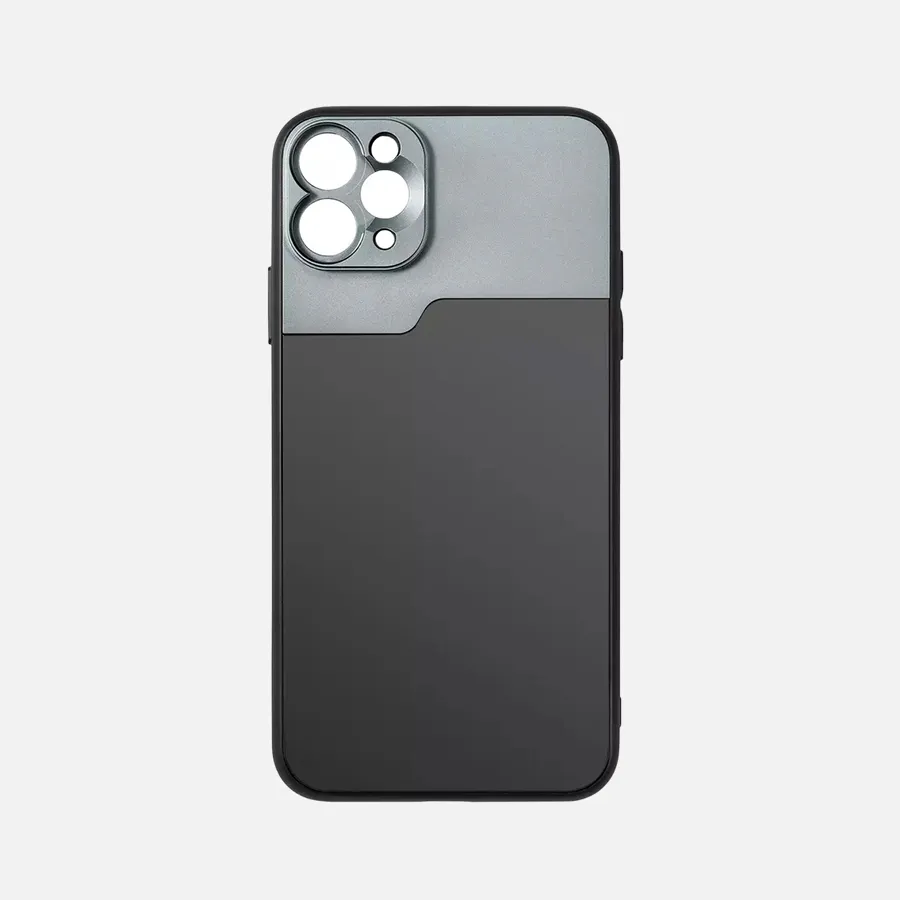
China Wholesale Dermoscopic Camera Factory Manufacturer Supplier - IBOOLO

2025 IBOOLO Dermatoscope Ultimate Guide: Technology, Product Selection & Clinical Applications

The New Era of Skin Health Monitoring

As we enter 2025, IBOOLO's innovative dermatoscope cameras are revolutionizing skin observation technology. Our complete product line - from the entry-level DE-400 to the advanced DE-4100 PRO - provides medical professionals with reliable tools for enhanced visualization of skin conditions. Each IBOOLO dermatoscope combines precision optics with user-friendly design, serving as valuable assistants in clinical practice while always requiring professional medical interpretation of findings.

What Makes IBOOLO Dermatoscopes Special?

IBOOLO dermatoscopes incorporate several proprietary technologies that set them apart:

  • Advanced Polarization System: Our patented dual-polarization technology provides optimal visualization for various skin types
  • High-Definition Optics: Multi-coated lenses deliver exceptional clarity with minimal distortion
  • Ergonomic Engineering: Carefully balanced designs reduce hand fatigue during extended use
  • Modular Design: Flexible configuration options to meet different clinical needs

Complete IBOOLO Product Line Overview

Our 2025 dermatoscope series offers solutions for every professional need:

Model Price Key Specifications Ideal Usage
DE-4100 PRO $799 30x magnification, multi-spectral imaging, 8GB internal storage Specialized dermatology clinics, research applications
DE-4100 $699 30x magnification, adaptive lighting system, magnesium alloy body High-volume clinical practice
DE-3100 PRO $599 20x magnification, fog-resistant lenses, IP54 protection Mobile medical professionals
DE-3100 $499 20x magnification, dual polarization modes General medical practice
DE-400 $179 10x magnification, ring LED illumination Basic screening, educational use

Selecting The Right IBOOLO Dermatoscope

Follow this professional guidance to choose your ideal model:

  1. Assess Your Primary Applications:
    • Routine skin checks: DE-3100 series
    • Detailed dermatological examination: DE-4100 series
    • Training and education: DE-400
  2. Consider Your Work Environment:
    • Clinic-based practice: DE-4100 models
    • Mobile/field work: DE-3100 PRO
  3. Evaluate Your Budget:
    • Cost-effective entry: DE-400
    • Best overall value: DE-3100
    • Premium features: DE-4100 PRO

Professional Features of IBOOLO PRO Series

Our PRO models offer enhanced capabilities for demanding clinical environments:

  • Rugged Construction:
    • Impact-resistant materials
    • Weather-sealed components
  • Advanced Imaging:
    • Multi-spectral analysis (DE-4100 PRO)
    • Anti-fog optical surfaces
  • Workflow Integration:
    • Seamless DICOM compatibility
    • One-button image capture

Maintaining Your IBOOLO Dermatoscope

Proper care ensures peak performance and longevity:

  • Daily Care:
    • Gently clean lenses with included microfiber cloth
    • Disinfect contact surfaces after each patient
  • Weekly Checks:
    • Inspect battery status
    • Verify all controls function properly
  • Monthly Maintenance:
    • Perform complete calibration
    • Install any available software updates

The IBOOLO Advantage

Choosing IBOOLO means investing in quality and support:

  • Optical Excellence: Precision-engineered lens systems
  • Proven Reliability: 98% operational uptime in clinical settings
  • Comprehensive Support:
    • Extended 3-year warranty on PRO models
    • Free lifetime software enhancements
    • Global service network

All IBOOLO dermatoscopes are designed as professional observation aids to support, not replace, qualified medical judgment.

People May Ask

What Are The Potential Drawbacks Or Limitations of Using Polarized Sunglasses?

The relentless alteration of light induced by polarized lenses can induce eye fatigue during the prolonged usage of technological devices, including smartphones, tablets, and laptops. This may present difficulties in conducting tasks, thereby rendering it an unfeasible choice for individuals whose occupation necessitates constant engagement with laptop screens.

Can Prizm Outperform Polarized?

Prizm lenses enhance your vision, while polarized lenses reduce glare from reflected light. Nevertheless, 100% UV protection from the sun's rays is provided by the majority of polarized and all Prizm lenses, preventing eye strain and burns when you're outside.

Who Are Competitors of DermTech?

Enzo Biochem (ENZ), Exagen (XGN), Centogene (CNTG), Akumin (AKUMQ), Renalytix (RNLX), Psychemedics (PMD), Personalis (PSNL), MDxHealth (MDXH), Genetron (GTH), and HH&L Acquisition (HHLA) are some of DermTech's primary rivals. The "medical laboratories" sector includes all of these businesses.

To What Extent Is The Dermlite Dermatoscope Magnified?

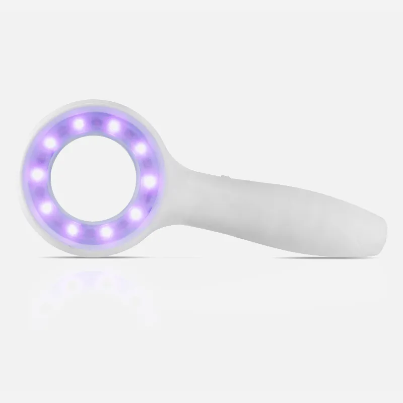
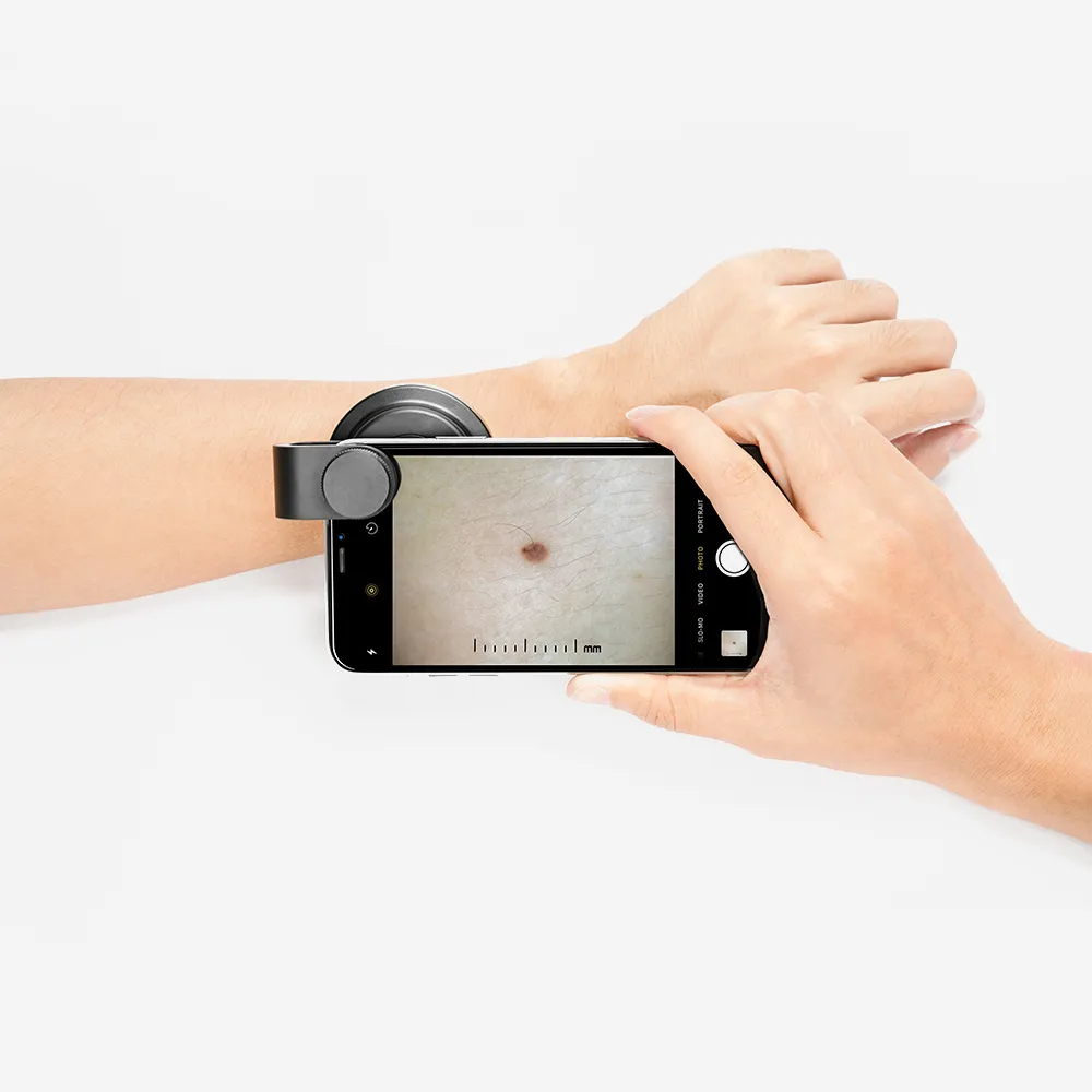
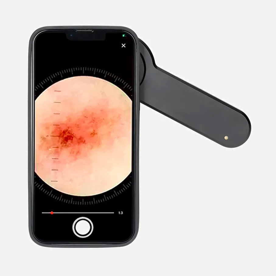
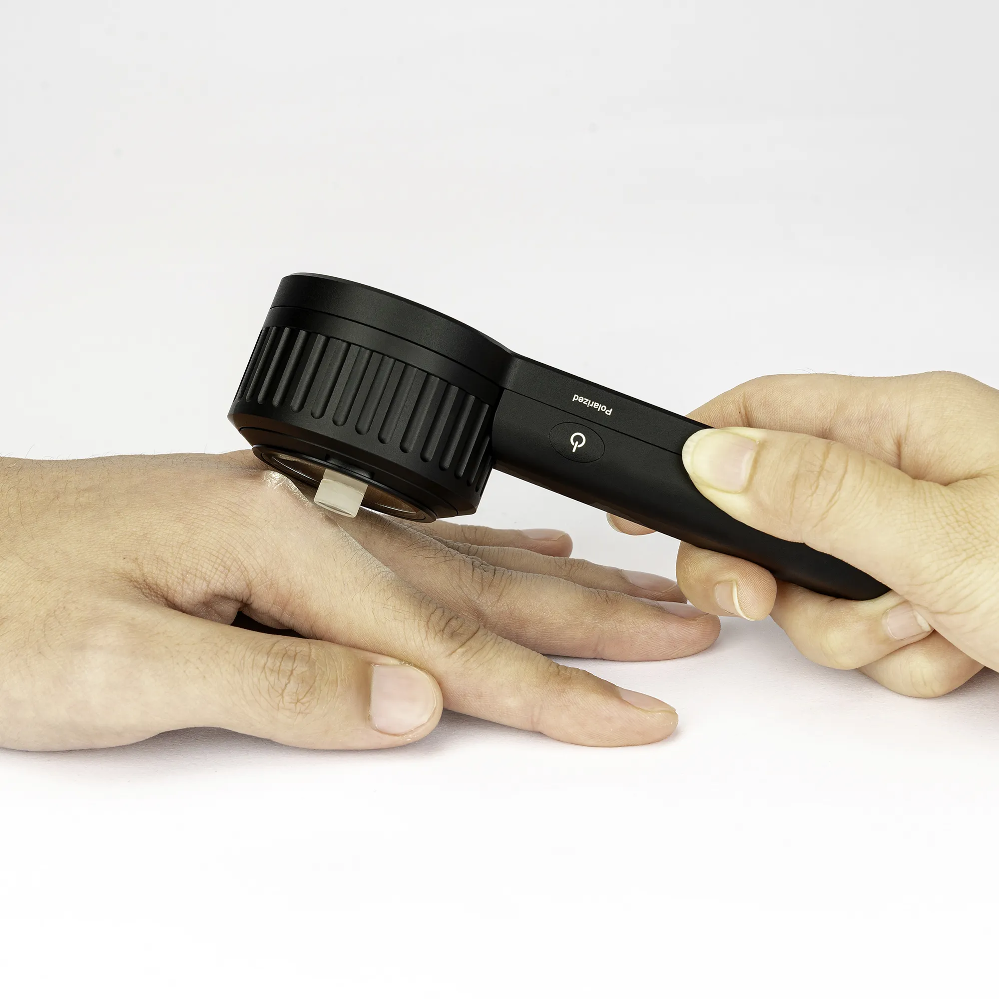
Handheld dermatoscope including changeable polarization, dermoscopic white and UV, torch LED, and 10x magnification. desktop charging base with auxiliary USB output and IceCap storage. 2 meter USB-C to USB-USB cable.

What Sort of Microscope Do Dermatologists Use?

Describe the dermatoscope. A dermatoscope is a light-powered magnifying instrument that is roughly the size of a tiny computer mouse that allows for a sharper, larger image of a skin lesion. Dermoscopy is the process of using a dermatoscope.

Which Four Categories of Skin Biopsies Exist?

What varieties of skin biopsies exist?Instead,Punch biopsy: The biopsy punch is a device used to retrieve a tiny, round sample of skin.Instead,Shave biopsy: A razor blade is used by your healthcare practitioner to remove a small portion of skin.Instead,Excisional biopsy: A scalpel is used by your healthcare professional to take a sample of skin.Instead,

How Much Should Be Paid for A Skin Biopsy?

Without insurance, a skin biopsy typically costs between $120 and $450. You may be charged an additional $50 to $350 for a lab evaluation. Is it necessary to perform the biopsy even if we believe we know what I have? Your skin's medical condition will determine all of this.

Which Metal Is The Weakest in The World?

Being a liquid at normal temperature, mercury has the lowest strength. With densities of 22.4 g/cc, gallium and iridium have the lowest densities. With a strength of 1510 megapascals apiece, tungsten and osmium are the strongest.

What Is The Best Biopsy?

biopsy guided by images.A needle biopsy is combined with an imaging technique, such as an MRI, CT scan, or ultrasound, in an image-guided biopsy. Your doctor can access suspicious areas, like the liver, lung, or prostate, that are not palpable through the skin with an image-guided biopsy.

What Are The Finest Dermoscopy Features for Bowen's Disease?

Findings: Glomerular vessels (90%) and a scaly surface (90%) were the most common dermoscopic patterns seen in most cases of BD. Furthermore, in pigmented BD, there were discovered to be 90% of minute brown globules regularly packed in a patchy distribution and 80% of structureless grey to brown pigmentation.

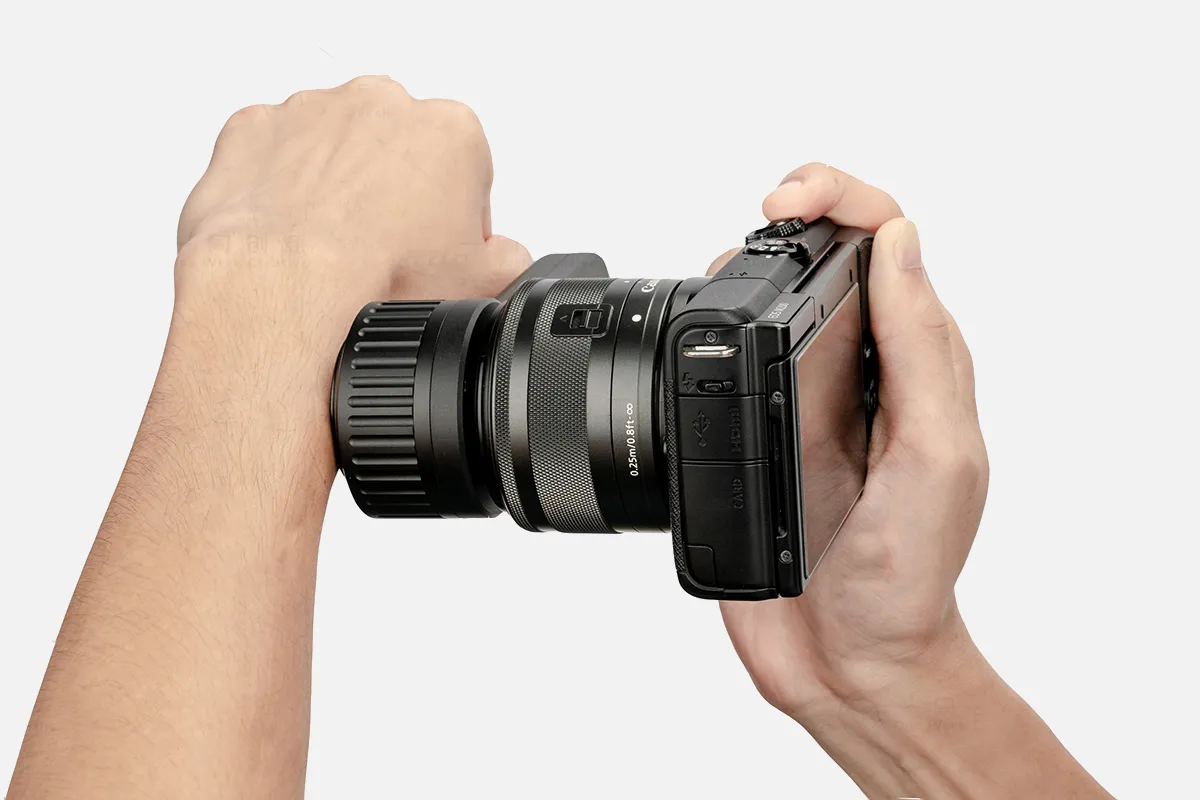
Dermoscopic Camera Products

OdontoMed2011 Gray DERMATOSCOPE Free CASE ODM Dermatology Examination

The MicroMax LED-lighted 60x-75x Carson Pocket Microscope (MM-200) and the Pocket Micro 20x-60x LED-lighted Zoom Field Microscope with Aspheric Lens System (MM-450) are both available in blue.

Utilizing a conventional microscope, the Dino-Lite USB Eyepiece Camera AM7025X - 5MP

Celestron Deluxe Handheld Digital Microscope (44302-C), Grey, Capture Your Discoveries

USB 2MP 数字显微敜 Q-Scope QS.20200-P

12 inch color HD portable video magnifier, Eschenbach Visolux Digital XL FHD

可互捚敜头手持数字显微敜放大镜 Supereyes B011 5.0MP 500X

UKCOCO Tiny Camera Digital Microscope Pocket Microscope Dermatoscope Microscope for Pocket Magnifying Glass Phone for Hand Led Phone Microscope Portable White Small Camera

Supereyes 11mm Diameter, B007, USB Digital Microscope Camera with 300X Zoom, Handheld Endoscope, Portable Magnifier Otoscope, and IP67 Waterproof for Windows, Mac, and Linux

Low Vision Aid: Bierley Maggie 5 Portable Ultra Slim Electronic Magnifier

Top Reviews

Linda

For my mother, who has macular degeneration, this has been incredibly beneficial. She can operate it rather easily, albeit it is a little tricky to move the object beneath the magnifier.

J. Socrates

While the Eschenbach device works perfectly, two out of the three light panels are no longer functional. The device's spoken voice says something extremely quickly. The sound is a little weak, but it seems to be saying "Battery low." So perhaps there's a problem with the lights' charging. I will investigate that. If this is the case, then another example of Eschenbach's clever design is to only turn on the main light when the battery becomes low rather than leaving the user in the dark. I'll follow up.

Hookedon

In the world of work, tools such as these are essential for someone who is legally blind and has low vision. One issue is that they can be quite expensive. I was looking for something that was lightweight and easy to carry. It's fantastic. I was worried about the screen breaking after watching YouTube videos and reading the IBOOLO description, so I thought I would have to buy a case. Its inclusion of a hard case astounded me. There are tons of features. It's simple to use the menus. Everybody talks. It has a 22X zoom. I usually use 12X for it. It's convenient that I can use the touch screen or the zoom buttons. The photos have excellent quality. clear and concise The picture on my other video magnifier, a 7-inch Optelec that is meant to be color, appears washed out. This gadget is quite excellent. I therefore adore this video magnifier. will utilize it daily at work. But there are two problems. I don't mean to be critical, but these wouldn't be a deal-breaker. I do, however, want the company to see this.

0

Have questions on gear or your order?

Our Gear Guides are here to help! Get personal advice from pro creatives

Name
Subject
Email address
How can we help?

Instant Answers

How Can Dermoscopy Images Be Captured?

Dermoscopy images can be captured and stored in different ways, such as: • Using a smartphone or tablet with dermoscopic adapter, which consisted in the package.• Using a digital camera

Dermoscopy images can be captured and stored in different ways, such as:

• Using a smartphone or tablet with dermoscopic adapter, which consisted in the package.
• Using a digital camera with dermoscopic adapter, there’s 49mm screw size camera adapter available to order now.

Compatible phone/tablet models:
All iPhone models, 95% Android phones, 90% tablet. For phone/tablet size in 5.25-14mm

Compatible camera models:
All camera with built 49mm filter screw, such as Canon EOS 70D, 80D, 90D; Canon EOS R7, R10, R50, R100; Canon M100, M200, M50, Mark II; Canon G7X Mark III, Sony ZV-1

How Can I Connect My Phone to My Dermatoscope?

There’s universal phone adapter for all our dermoscopes. Please check the installation procedure bellow or watch operation guide. Smartphone Connector (1) Place phone adapter screw in the center of smartphone’s

There’s universal phone adapter for all our dermoscopes. Please check the installation procedure bellow or watch operation guide.

Smartphone Connector

(1) Place phone adapter screw in the center of smartphone’s main camera.
(2) Screw magnet attachment on phone adapter.
(3) Put dermoscope’s back ring and magnet attachment together

Take The Best Images

You need to adjust the focus ring after the dermoscpe connected on smartphone to get the best images.

How Can I Clean My Dermoscopy after Usage?

Cleaning your dermoscopy after usage is important to prevent cross-contamination and infection. The cleaning method may vary depending on the type and model of your dermoscopy, so you should always

Cleaning your dermoscopy after usage is important to prevent cross-contamination and infection. The cleaning method may vary depending on the type and model of your dermoscopy, so you should always follow the manufacturer’s instructions. However, some general steps are:

• Turn off and disconnect your dermoscopy from any power source or device.

• Wipe off any visible dirt or debris from the dermoscopy with a soft cloth or tissue.

• Disinfect the dermoscopy with an alcohol-based wipe or spray, or a disinfectant solution recommended by the manufacturer. Make sure to cover all surfaces, especially the lens and contact plate.

• Let the dermoscopy air dry completely before storing it in a clean and dry place.

• Do not use abrasive or corrosive cleaners, solvents, or detergents that may damage the dermoscopy.

• Do not immerse the dermoscopy in water or any liquid, unless it is waterproof and designed for immersion.

You should clean your dermoscopy after each use, or at least once a day if you use it frequently. You should also check your dermoscopy regularly for any signs of damage or malfunction, and contact the manufacturer or service provider if needed.

Polarized VS Non-Polarized Dermoscopy

A dermoscopy is a device that allows the examination of skin lesions with magnificationand illumination. By revealing subsurface structures and patterns that are not visible tothe naked eye. It can

A dermoscopy is a device that allows the examination of skin lesions with magnificationand illumination. By revealing subsurface structures and patterns that are not visible tothe naked eye. It can improve the diagnose accuracy of skin lesions, such as melanoma,basal cell carcinoma, seborrheic keratosis, etc.

There are two main types of dermoscopy: Non polarized and polarized dermoscopy.We’ve fitted most of our dermoscopys with polarized and non-polarized light. They canbe used in multiple skin structures.

Non-polarized contact Mode

In non-polarized mode, the instrument can provide information about the superficialskin structures, such as milia-like cysts, comedo-like openings, and pigment in theepidemis.

The dermoscopy requires applying a liquid such as mineral oil or alcohol to the skin andplacing the lens in contact with the skin. This reduces surface reflection and enhancesthe view of subsurface structures.

Image with non-polarized light (DE-3100)

Polarized contact Mode

In polarized mode, the instrument allows for visualization for deeper skin structures,such as blood vessels, collagen, and pigment in the dermis.

The dermoscopy does not need to be in contact with the skin or use any liquid. Theirpolarized light can help to eliminate surface reflection and allow visualization ofvascular structures.

Image with polarized light (DE-3100)

Polarized non-contact Mode

The dermoscopy can also use polarized light to examine the skin without direct contact.

In polarized non-contact mode, the instrument allows for examination infected areasand lesions that are painful for the patient, or the difficult to contact pigmented lesions,such as nails and narrow areas.

The contact plate should be removed in this mode, and it does not require applying aliquid to the skin. As it doesn’t require pressure or fluid application on the skin, it canalso avoid cross-contamination and infection risk.

Image in polarized non-contact mode (DE-3100)

How Effectiveness Is Dermoscopy

Compared with visual inspection, the dermoscopy can be used to capture and store skin lesion photos, which play an important role in early skin cancer examination. The dermoscopy allows the

Compared with visual inspection, the dermoscopy can be used to capture and store skin lesion photos, which play an important role in early skin cancer examination.

The dermoscopy allows the examination of skin lesions with magnification and illumination. This can be greatly avoiding the factors that cause interference to visual detection. Such as lighting, skin color, hair and cosmetics.

Several studies have demonstrated that dermoscopy is useful in the identification of melanoma, when used by a trained professional.

It may improve the accuracy of clinical diagnosis by up to 35%
It may reduce the number of harmless lesions that are removed
In primary care, it may increase the referral of more worrisome lesions and reduce the referral of more trivial ones

A 2018 Cochrane meta-analysis published the accuracy of dermoscopy in the detection.

Table 1. Accuracy of dermoscopy in the detection of melanoma in adults
Detection Method Sensitivity, % Specificity, % Positive Likelihood Ratio NegativeLikelihood Ratio
Visual inspection alone (in person) 76 75 3.04 0.32
Dermoscopy with visual inspection (in person) 92 95 18 0.08
Image-based visual inspection alone (not in person) 47 42 0.81 1.3
Dermoscopy with image-based visual inspection (not in person) 81 82 4.5 0.23
ROC—receiver operating characteristic. *Estimated sensitivity calculated on the summary ROC curve at a fixed specificity of 80%.

As we can see, the dermoscope can improve the accuracy of diagnosis of skin lesions, especially melanoma.

Table 1. Accuracy of dermoscopy in the detection of melanoma in adults
Detection Method Sensitivity, % Specificity, % Positive Likelihood Ratio NegativeLikelihood Ratio
Visual inspection alone (in person) 79 77 3.4 0.27
Dermoscopy with visual inspection (in person) 93 99 93 0.07
Image-based visual inspection alone (not in person) 85 87 6.5 0.17
Dermoscopy with image-based visual inspection (not in person) 93 96 23 0.07
ROC—receiver operating characteristic. *Estimated sensitivity calculated on the summary ROC curve at a fixed specificity of 80%.

Characteristics of the dermatoscopic structure of the skin lesions include:

• Symmetry or asymmetry
• Homogeny/uniformity (sameness) or heterogeny (structural differences across the lesion)
• Distribution of pigment: brown lines, dots, clods and structureless areas
• Skin surface keratin: small white cysts, crypts, fissures
• Vascular morphology and pattern: regular or irregular
• Border of the lesion: fading, sharply cut off or radial streaks
• Presence of ulceration

There are specific dermoscopic patterns that aid in the diagnosis of the following pigmented skin lesions:

• Melanoma
• Moles (benign melanocytic naevus)
• Freckles (lentigos)
• Atypical naevi
• Blue naevi
• Seborrhoeic keratosis
• Pigmented basal cell carcinoma
• Haemangioma

No products in the cart.

No products in the cart.

Hot Search Terms