


Article
Something About IBOOLO
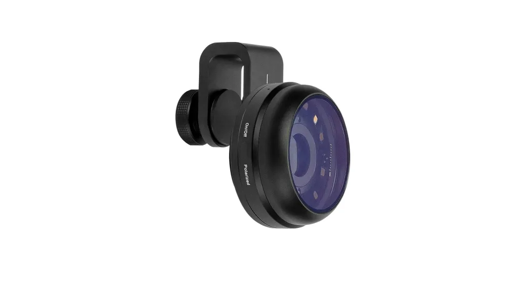
Why did IBOOLO start developing dermatoscopes? In 2012, IBOOLO was born from a simple dream: to change people’s lives with optical technology. Initially focused on smartphone lens attachments, we earned global acclaim for our superior imaging. But a casual conversation redirected our path. A German user, amazed by our lenses, exclaimed: “Your optics are phenomenal!…
Why did IBOOLO start developing dermatoscopes?
In 2012, IBOOLO was born from a simple dream: to change people’s lives with optical technology. Initially focused on smartphone lens attachments, we earned global acclaim for our superior imaging. But a casual conversation redirected our path. A German user, amazed by our lenses, exclaimed: “Your optics are phenomenal! Why not create dermatoscopes? Doctors here desperately need tools like this.
This comment deeply moved us. We began to look into the dermatoscope market and discovered a shocking reality: dermatoscopes on the market were extremely expensive, often costing over a thousand dollars, while the number of skin cancer patients worldwide was huge. This made us realize that a dermatoscope is not just a tool but a hope for countless people’s health. So, we decided to invest all our optical technology expertise into dermatoscope development. We visited hospitals and clinics, listened to doctors’ needs, and repeatedly refined the product design. Our goal was clear: to create a dermatoscope that is both affordable and high-performance, allowing every doctor in need to have access to it.
When did IBOOLO launch its first dermatoscope?
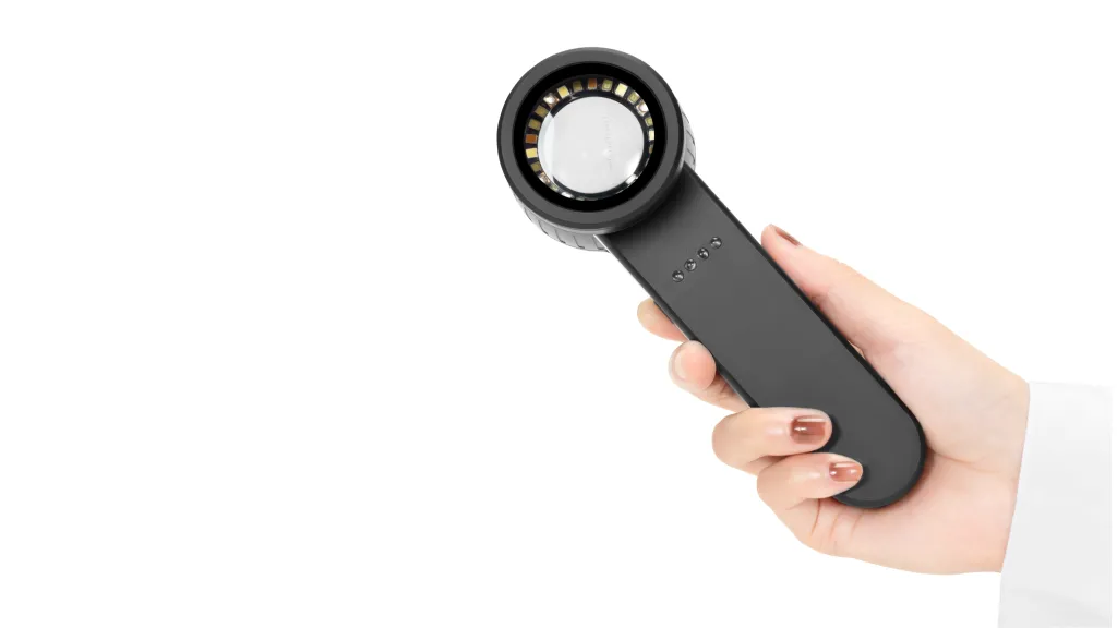
Five years ago, our first dermatoscope, the DE-200, was officially launched. It not only inherited our core optical technology but also significantly reduced costs through innovative design. The product received positive feedback from global users as soon as it was released. This fueled our resolve: to make skin health achievable through technology. Since then, we’ve evolved relentlessly—launching pocket-sized DE-300, DE-400, and handheld DE-3100, DE-4100. We will continue to launch more and better dermatoscopes in the future. We believe that a dermatoscope is not just a device but a bridge connecting doctors and patients, an important tool for improving global skin health.
What new dermatoscope products will IBOOLO release next?
Currently, IBOOLO’s latest pocket-sized dermatoscope, the DE-500, and the latest handheld dermatoscope, the DE-5100, will be launched in 2025. Compared to the DE-400 and DE-4100, these two models use more advanced manufacturing processes and have improved functionality. Furthermore, IBOOLO will not be limited to optical dermatoscopes. We are already preparing to launch a digital dermatoscope series, including portable and desktop models. In the near future. IBOOLO’s future products will expand into even broader areas. We are also developing and manufacturing otoscopes, eye fundus cameras, ophthalmoscopes, anterior scopes, and more.
Who are the optical designers behind IBOOLO Dermatoscope?
One of the core competencies of IBOOLO dermatoscope lies in the strong team of optical designers behind it. This team consists of senior optical experts from Taiwan and Hong Kong, who have accumulated more than 20 years of optical design experience in world-renowned camera companies. IBOOLO designers have migrated these cutting-edge technologies into the development of the dermatoscope. In particular, they have demonstrated their deep professional expertise in the combination and matching of optical glass and the application of polarised light technology.

In addition to the local designer team, IBOOLO has also invited a team of German optical consultants to provide technical support. Germany is a global leader in the field of optics, especially in the design of optical systems for medical devices. The addition of the German consultant team not only helped IBOOLO to solve a number of technical problems, but also ensured that the product design meets the international leading standards.
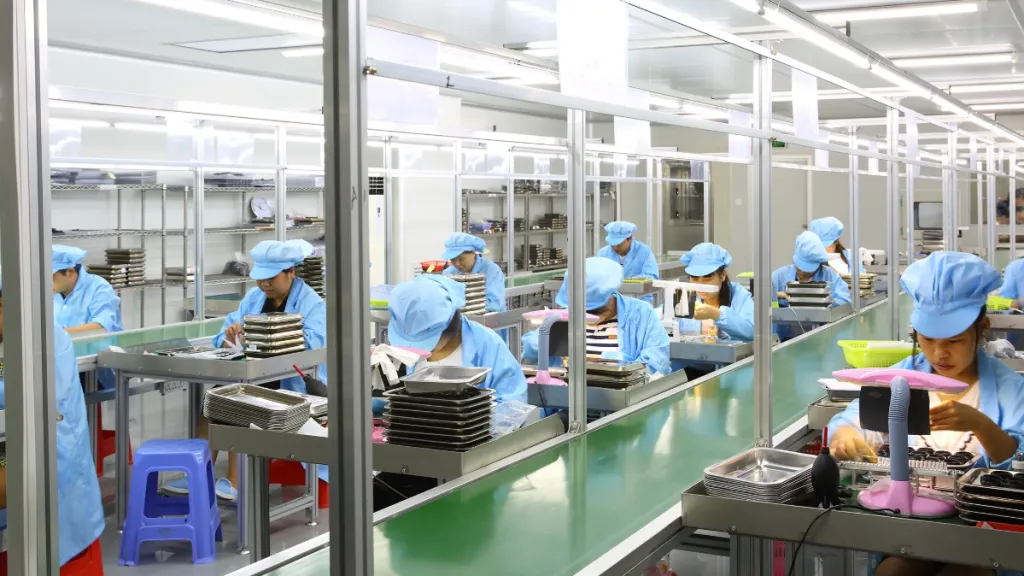
How are IBOOLO dermatoscopes produced?
Every dermatoscope from IBOOLO starts in a highly clean and dust-free workshop. The assembly of optical components is one of the most critical aspects in the production of dermatoscopes. IBOOLO employs fully automated precision equipment for the cleaning, coating and assembly of optical glass to ensure that each lens meets the precision required by the design. During the production process, IBOOLO also pays special attention to the aging test of the equipment. Before leaving the factory, each dermatoscope is subjected to more than 500 hours of aging tests, including high temperature, high humidity, low temperature and many other extreme environments. There is also an optical performance test, which examines the device’s imaging resolution, colour reproduction and polarised light effect, to ensure that every dermatoscope delivers clear, accurate images.
What defines IBOOLO’s philosophy?
Technology for the benefit of all, protecting global skin health.
Through continuous technological innovation and cost optimization, we are committed to ensuring that every doctor and patient can use high-quality dermatoscopes. From smartphone lenses to dermatoscopes, user needs drive us. We listen to doctors’ voices, we focus on patients’ pain points, we solve real problems with our products. Skin diseases are a global health challenge, and we hope to raise awareness of skin health by popularizing dermatoscope technology.
What is IBOOLO’s vision?
In the future, IBOOLO will continue to delve deeper into optical technology and promote the popularization and upgrading of dermatoscopes. We hope that through our efforts, more doctors will have access to high-quality dermatoscopes, and more patients will receive timely diagnoses and treatments.


