IBOOLO Dermatoscope Accessories – IBOOLO

The headquarters of Shenzhen Iboolo Optics Co.Ltd are in Shenzhen, Guangdong. We are specialized in the R&D, production, and marketing of all varieties of Macro lens, Woods Lamp, Dermatoscope and Microscope.

Article

IBOOLO Dermatoscope Accessories

What Are the Available Accessories for IBOOLO Dermatoscopes? When it comes to enhancing the functionality of your IBOOLO dermatoscope, there are several accessories available to suit different needs. These include the small contact plate, eye piece, phone case, and additional components like the contact plate and back-up battery. These accessories are designed to improve your…

What Are the Available Accessories for IBOOLO Dermatoscopes?

When it comes to enhancing the functionality of your IBOOLO dermatoscope, there are several accessories available to suit different needs. These include the small contact plate, eye piece, phone case, and additional components like the contact plate and back-up battery. These accessories are designed to improve your dermatoscopic experience, whether you’re a professional dermatologist or a medical student. Let’s dive into each accessory and explore how they can benefit your practice.

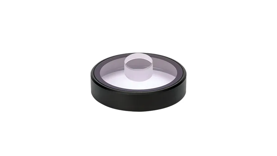
Why Should You Use a Small Contact Plate?

The small contact plate is a crucial accessory for IBOOLO dermatoscopes, especially when examining hard-to-reach areas like the spaces between fingers and toes, the nasal wings, or the ear canal. With a diameter of 9mm, this compact plate is perfect for detailed dermatoscopic examinations in confined spaces. It allows for better visibility and accuracy when assessing lesions such as pigmented moles, plantar warts, or even scabies infestations. The small contact plate is compatible with IBOOLO models like the DE-4100, DE-3100, DE-4100 PRO, and DE-3100 PRO, making it a versatile addition to your dermatoscope setup. This accessory is particularly useful for examining lesions in interdigital areas, where standard contact plates may not fit comfortably.

IBOOLO Small Contact Plate

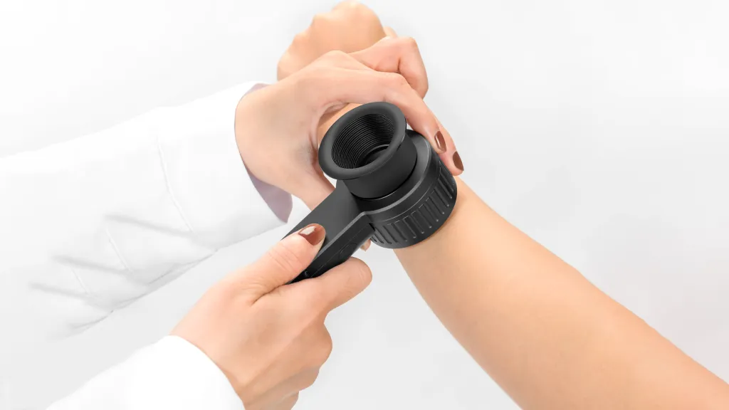
How Does the Eye Piece Enhance Your View?

The eye piece is another essential accessory for IBOOLO dermatoscopes. Made from high-quality aluminum, this durable component attaches securely to your dermatoscope via a magnetic connection. The eye piece helps eliminate unwanted reflections caused by overhead lights or other light sources, ensuring a clear and uninterrupted view of the skin. Whether you’re examining pigmented lesions or other dermatological conditions, the eye piece provides a comfortable and stable viewing experience, enhancing both precision and comfort during your assessments. This accessory is particularly beneficial in environments with strong ambient light, where reflections might otherwise obscure critical details.

IBOOLO Eye Piece
IBOOLO Eye Piece

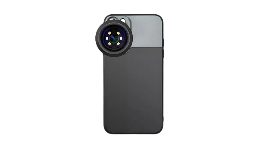
Can a Phone Case Make Using Your Dermatoscope Easier?

Absolutely! IBOOLO offers specialized phone cases designed to simplify the process of connecting your dermatoscope to your smartphone. While the standard IBOOLO dermatoscope kit includes a universal phone adapter and magnetic ring, these accessories can sometimes be cumbersome to carry around. By investing in an IBOOLO phone case, you can enjoy hassle-free connectivity between your dermatoscope and phone. The phone case not only protects your device during daily use but also ensures a seamless connection for capturing images or videos during dermatoscopic examinations. Currently, IBOOLO offers phone cases for Apple and Samsung devices, with more models set to be released in the future. This accessory is ideal for practitioners who frequently use their dermatoscope for documentation or telemedicine purposes. For example, the Pocket Dermatoscope series, DE-300 and DE-400 can even be rotated to connect to a cell phone directly through the threaded design on the case.

IBOOLO Phone Case
IBOOLO Phone Case

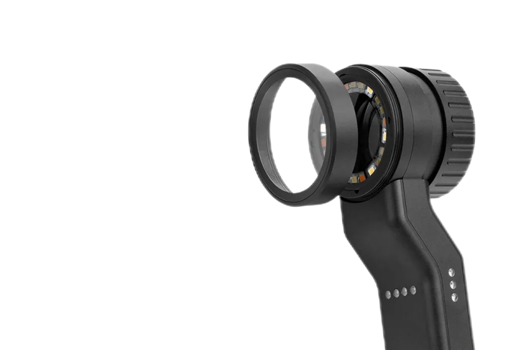
Is It Easy to Replace Damaged Components?

Yes, replacing damaged components of your IBOOLO dermatoscope is straightforward. If you accidentally drop your device or expose it to water, components like the contact plate or battery can be easily purchased and replaced. IBOOLO provides these spare parts on their official website, and their customer support team is available to guide you through the replacement process. Whether you need a new contact plate or a back-up battery, these accessories ensure that your device remains functional and reliable, even in challenging situations. This is particularly important for maintaining the longevity and performance of your dermatoscope, especially in clinical settings where equipment reliability is critical.

IBOOLO Contact Plate

What new accessory is next for IBOOLO?

IBOOLO is pleased to announce the upcoming release of disposable contact plates for dermoscopy. These disposable contact plates are designed for situations where there is a risk of infectious skin infection or disease. The disposable contact plates are single-use and maximize hygiene and safety during the examination process. For example, when a physician needs to use a dermatoscope to visualize skin lesions on a private and infected area such as the genitals or anus, the disposable contact plate can be used. This new accessory will soon be available for ordering through IBOOLO’s official channels, providing a convenient solution for practitioners concerned with infection control.

Conclusion

IBOOLO dermatoscope accessories are designed to enhance your dermatological practice by providing convenience, accuracy, and durability. From the small contact plate for detailed examinations to the eye piece for clearer views, and the phone case for hassle-free connectivity, these accessories cater to a wide range of needs. Additionally, the availability of replacement components like contact plates and batteries ensures that your device remains in optimal condition. Whether you’re a professional or a student, investing in these accessories can significantly improve your dermatoscopic experience.

Tags

Share this article

0

No products in the cart.

Have questions on gear or your order?

Our Gear Guides are here to help! Get personal advice from pro creatives

Name
Subject
Email address
How can we help?

Instant Answers

How can dermoscopy images be captured?

Dermoscopy images can be captured and stored in different ways, such as: • Using a smartphone or tablet with dermoscopic adapter, which consisted in the package.• Using a digital camera

Dermoscopy images can be captured and stored in different ways, such as:

• Using a smartphone or tablet with dermoscopic adapter, which consisted in the package.
• Using a digital camera with dermoscopic adapter, there’s 49mm screw size camera adapter available to order now.

Compatible phone/tablet models:
All iPhone models, 95% Android phones, 90% tablet. For phone/tablet size in 5.25-14mm

Compatible camera models:
All camera with built 49mm filter screw, such as Canon EOS 70D, 80D, 90D; Canon EOS R7, R10, R50, R100; Canon M100, M200, M50, Mark II; Canon G7X Mark III, Sony ZV-1

How can I connect my phone to my dermatoscope?

There’s universal phone adapter for all our dermoscopes. Please check the installation procedure bellow or watch operation guide. Smartphone Connector (1) Place phone adapter screw in the center of smartphone’s

There’s universal phone adapter for all our dermoscopes. Please check the installation procedure bellow or watch operation guide.

Smartphone Connector

(1) Place phone adapter screw in the center of smartphone’s main camera.
(2) Screw magnet attachment on phone adapter.
(3) Put dermoscope’s back ring and magnet attachment together

Take The Best Images

You need to adjust the focus ring after the dermoscpe connected on smartphone to get the best images.

How can I clean my dermoscopy after usage?

Cleaning your dermoscopy after usage is important to prevent cross-contamination and infection. The cleaning method may vary depending on the type and model of your dermoscopy, so you should always

Cleaning your dermoscopy after usage is important to prevent cross-contamination and infection. The cleaning method may vary depending on the type and model of your dermoscopy, so you should always follow the manufacturer’s instructions. However, some general steps are:

• Turn off and disconnect your dermoscopy from any power source or device.

• Wipe off any visible dirt or debris from the dermoscopy with a soft cloth or tissue.

• Disinfect the dermoscopy with an alcohol-based wipe or spray, or a disinfectant solution recommended by the manufacturer. Make sure to cover all surfaces, especially the lens and contact plate.

• Let the dermoscopy air dry completely before storing it in a clean and dry place.

• Do not use abrasive or corrosive cleaners, solvents, or detergents that may damage the dermoscopy.

• Do not immerse the dermoscopy in water or any liquid, unless it is waterproof and designed for immersion.

You should clean your dermoscopy after each use, or at least once a day if you use it frequently. You should also check your dermoscopy regularly for any signs of damage or malfunction, and contact the manufacturer or service provider if needed.

Polarized VS Non-polarized Dermoscopy

A dermoscopy is a device that allows the examination of skin lesions with magnificationand illumination. By revealing subsurface structures and patterns that are not visible tothe naked eye. It can

A dermoscopy is a device that allows the examination of skin lesions with magnificationand illumination. By revealing subsurface structures and patterns that are not visible tothe naked eye. It can improve the diagnose accuracy of skin lesions, such as melanoma,basal cell carcinoma, seborrheic keratosis, etc.

There are two main types of dermoscopy: Non polarized and polarized dermoscopy.We’ve fitted most of our dermoscopys with polarized and non-polarized light. They canbe used in multiple skin structures.

Non-polarized contact Mode

In non-polarized mode, the instrument can provide information about the superficialskin structures, such as milia-like cysts, comedo-like openings, and pigment in theepidemis.

The dermoscopy requires applying a liquid such as mineral oil or alcohol to the skin andplacing the lens in contact with the skin. This reduces surface reflection and enhancesthe view of subsurface structures.

Image with non-polarized light (DE-3100)

Polarized contact Mode

In polarized mode, the instrument allows for visualization for deeper skin structures,such as blood vessels, collagen, and pigment in the dermis.

The dermoscopy does not need to be in contact with the skin or use any liquid. Theirpolarized light can help to eliminate surface reflection and allow visualization ofvascular structures.

Image with polarized light (DE-3100)

Polarized non-contact Mode

The dermoscopy can also use polarized light to examine the skin without direct contact.

In polarized non-contact mode, the instrument allows for examination infected areasand lesions that are painful for the patient, or the difficult to contact pigmented lesions,such as nails and narrow areas.

The contact plate should be removed in this mode, and it does not require applying aliquid to the skin. As it doesn’t require pressure or fluid application on the skin, it canalso avoid cross-contamination and infection risk.

Image in polarized non-contact mode (DE-3100)

How effectiveness is dermoscopy

Compared with visual inspection, the dermoscopy can be used to capture and store skin lesion photos, which play an important role in early skin cancer examination. The dermoscopy allows the

Compared with visual inspection, the dermoscopy can be used to capture and store skin lesion photos, which play an important role in early skin cancer examination.

The dermoscopy allows the examination of skin lesions with magnification and illumination. This can be greatly avoiding the factors that cause interference to visual detection. Such as lighting, skin color, hair and cosmetics.

Several studies have demonstrated that dermoscopy is useful in the identification of melanoma, when used by a trained professional.

It may improve the accuracy of clinical diagnosis by up to 35%
It may reduce the number of harmless lesions that are removed
In primary care, it may increase the referral of more worrisome lesions and reduce the referral of more trivial ones

A 2018 Cochrane meta-analysis published the accuracy of dermoscopy in the detection.

Table 1. Accuracy of dermoscopy in the detection of melanoma in adults
Detection Method Sensitivity, % Specificity, % Positive Likelihood Ratio NegativeLikelihood Ratio
Visual inspection alone (in person) 76 75 3.04 0.32
Dermoscopy with visual inspection (in person) 92 95 18 0.08
Image-based visual inspection alone (not in person) 47 42 0.81 1.3
Dermoscopy with image-based visual inspection (not in person) 81 82 4.5 0.23
ROC—receiver operating characteristic. *Estimated sensitivity calculated on the summary ROC curve at a fixed specificity of 80%.

As we can see, the dermoscope can improve the accuracy of diagnosis of skin lesions, especially melanoma.

Table 1. Accuracy of dermoscopy in the detection of melanoma in adults
Detection Method Sensitivity, % Specificity, % Positive Likelihood Ratio NegativeLikelihood Ratio
Visual inspection alone (in person) 79 77 3.4 0.27
Dermoscopy with visual inspection (in person) 93 99 93 0.07
Image-based visual inspection alone (not in person) 85 87 6.5 0.17
Dermoscopy with image-based visual inspection (not in person) 93 96 23 0.07
ROC—receiver operating characteristic. *Estimated sensitivity calculated on the summary ROC curve at a fixed specificity of 80%.

Characteristics of the dermatoscopic structure of the skin lesions include:

• Symmetry or asymmetry
• Homogeny/uniformity (sameness) or heterogeny (structural differences across the lesion)
• Distribution of pigment: brown lines, dots, clods and structureless areas
• Skin surface keratin: small white cysts, crypts, fissures
• Vascular morphology and pattern: regular or irregular
• Border of the lesion: fading, sharply cut off or radial streaks
• Presence of ulceration

There are specific dermoscopic patterns that aid in the diagnosis of the following pigmented skin lesions:

• Melanoma
• Moles (benign melanocytic naevus)
• Freckles (lentigos)
• Atypical naevi
• Blue naevi
• Seborrhoeic keratosis
• Pigmented basal cell carcinoma
• Haemangioma

We use cookies on this website to provide a better user experience. By continuing to browse the website, you are giving your consent to receive cookies on this site. For more details please read our Privacy Policy.

Hot Search Terms