Article

Dermoscopy of Wart

Warts are noncancerous skin growths caused by the human papilloma virus (HPV). HPV infecting through the skin and mucous membranes enters to its target epithelial cells where proliferates in these polarized keratinocytes inducing abnormal cell growth, differentiation and warts formation. Dermoscopy provides a more detailed scrutiny of lesions than can be achieved with the naked eye…

Warts are noncancerous skin growths caused by the human papilloma virus (HPV). HPV infecting through the skin and mucous membranes enters to its target epithelial cells where proliferates in these polarized keratinocytes inducing abnormal cell growth, differentiation and warts formation. Dermoscopy provides a more detailed scrutiny of lesions than can be achieved with the naked eye and is based on cutting-edge optical technology, to better appreciate features visible at skin surface. This technique permits the detection of minor variations, from mild dystrophy to modest abnormalities within follicular framework which facilitate the recognition of compact wart-like lesions.

The Basics of Warts

According to their location and form, warts are mainly divided into common warts, plantar warts, flat warts and genital wars. Warts are most commonly passed through skin-to-skin, self-inoculation, sexual contact and perinatal transmission.

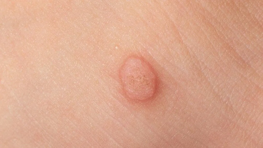
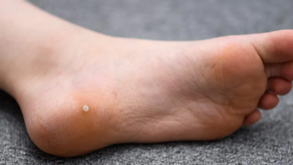
Common warts tend to be grayish-brown or brownish, slightly drooping (like a soybean), hard papillary growths with a rough surface. These are considered plantar warts based on the appearance of these typical circumscribed, keratinized, and rough-surfaced papules that appear as slightly elevated rings of hyperkeratosis. Flat warts often present as well-demarcated apical flat papules the size of grains of rice and sesame seeds. Genital warts are papillary or cauliflower-like projections that are susceptible to infection by secondary microbial erosion and pus, accompanied by itching and pain.

Flat Wart
Flat Wart

Unique Advantages of Dermoscopy in the Diagnosis of Warts

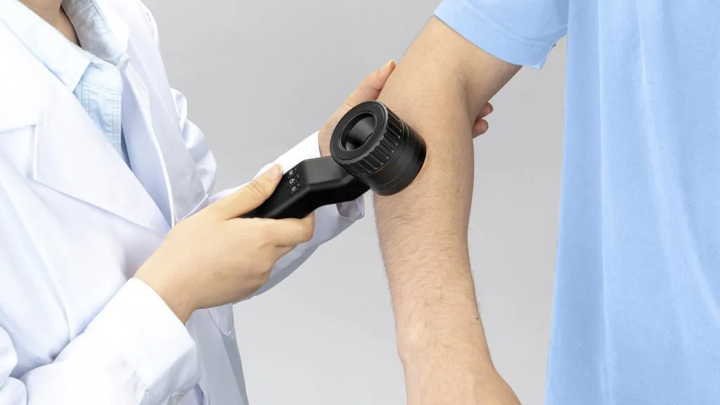
A dermatoscopic instrument employs the principles of optics to visualize surface morphology and texture. The features are seen in it. Dermoscopy magnifies the skin surface structure and is a procedure that physicians can use to more closely examine morphology, of colour variations in pigmentation distribution or colour asymmetry variegation on any other lesion site details for an accurate diagnosis. This increased detail allows for better detection and diagnosis of early or small skin lesions, which can be difficult to see with the naked eye.

Dermoscopy readily reveals the delicate modification in epidermal and hair follicle substructure of warts as hyperkeratosis, thickening of spinous layer over wart area which help doctors to diagnose accurately. Simultaneously, dermoscopy magnification and image enhancement effects can capture lots of wafer-like lesion very small in number difficult to be visualized.

Warts on Different Parts of the Body under Dermoscopy

Common Warts: Many areas showing papillomatous structures in a compact pattern. The frogspawn lesion will have a central red punctate/looped vessel with several fused vessels, usually  characterised as low to intermediate grade however occasionally having some ipsilateral associated punctate bleeding and peripheral adjacent blood lines that are present creating this appearance.

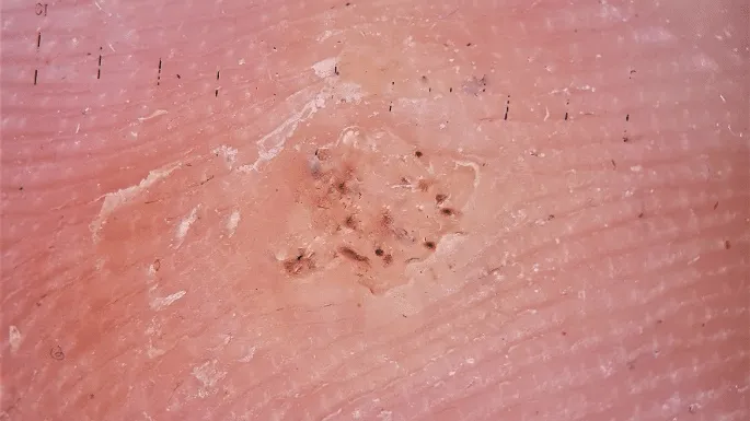
Plantar Warts: Displayed against a yellow unstructured background are variable reddish to dark brown dots or linear bleeding.

Flat Warts: Multiple papules, pinhead sized to 2.5mm in diameter with a hyperpigmented-to-violaceous appearance and peripheral white halo At times, punctate or even linear vessels might be observed.

Genital Warts: A mosaic pattern with papillary structures grown out can be observed

Plantar Wart
Plantar Wart

Warts and other skin lesions

Warts

Clinical Features: Verrucous architecture with an irregular papillomatous surface often likened to a cauliflower; location frequently on the extremities (hands and feet)

Dermoscopy: Presence of small, dark thrombosed capillaries or dots in conjunction with finger-like projections and a distinctive mosaic pattern Seborrheic Keratosis

Seborrheic Keratosis

Clinical Features: Waxlike appearance appearing to be stuck onto skin; colour brownade, black and or tan.

Dermoscopy: milia-like cystic structures, comedo-like openings, cerebriform pattern.

Nevus

Clinical Features: Characterized by small, darkly pigmented papules, which are predominantly located on the facial and cervical regions, with heightened prevalence in individuals possessing darker skin pigmentation.

Dermoscopy: Reveals a uniformly pigmented lesion with an unvascularized smooth cutaneous surface.

Acne

Clinical Features: Erythematous/Inflamed papules, pustular but also comedonal distribution; facial/dorsal/thoracic involvement

Dermoscopy: Presence of orifices related to hair follicles, peripheral erythematous areas and occasionally pustular lesions.

IBOOLO Dermoscopy
IBOOLO Dermoscopy

Dermoscopy

Dermoscopy is a non-invasive, in-vivo technique designed to identify morphologic structures on the skin not typically visible at submicroscopic levels that can help distinguish features such as color and vascular details of pigmented lesions. The discovery of these well-recognized patterns plays an important role in distinguishing among different dermatoses. For example, in pigmented skin lesions (pigmented nevi and melanomas), the ability of dermoscopy to distinguish between these two is clearly established. In addition, as a kind of non-invasive dermatological examination method, dermoscopy can alleviate the harm and pain to patients (at least relatively). Through dermoscopic observation and diagnosis, doctors can more accurately determine the nature and extent of skin lesions, thus avoiding unnecessary skin biopsies.

Dermoscopy in the Treatment of Warts

Warts are managed according to the dermoscopic images. This permitted stepwise observation of change in colour intensity and morphologic characteristics, as well vasculature appearance within the wart with treatment. So it leads to early detection of response to treatment and abnormality like noneffective regression or new onset.

Besides, dermoscopic assessment is an objective endpoint to judge the efficacy. Dermoscopic change in terms of area reduction as well as an improved vascular pattern, etc., are some specific changes seen which doctors use it to assess responses before and after therapy. Because of this, the number reduced patient visits which greatly improved accuracy and effectiveness care result decreased pain but increased significantly accurate.

Dermoscopy of Wart
Dermoscopy of Wart

How to Prevent Warts?

We should not touch another person’s warts in the first place, and secondly we need to be careful of sharing private things like cleaning tools or razors shoes, since touching transmits very easily because it is caused by human Papillomavirus. If you just used hand sanitizer and water to wash your hands immediately after contact with things or surfaces which might be contaminated with warts. Seconding is that try not to have skin contactin public places especially in areas which are wet like public toilets, swimming pools and gyms in order to prevent chances of getting a wart infection.

How to Treat Warts?

Wart treatment options include use of drugs, cryotherapy and laser therapy. Medication therapy mainly involves Flurouracil Ointment, Imiquimod Ointment, Salicylic Acid Ointment, and Tazarotene Creams which act as wart remover creams. These medications help dissolve wart tissues, limit replication of viral cells or destroy viral particles thus improving symptoms associated with warts. Cryotherapy is a technique for treating common warts through rapid freezing using liquid nitrogen. The low temperature of liquid nitrogen leads to freezing damage within the wart tissue; this destroys the virus particles as well as the wart cells hence treating it. Laser treatment removes warts by directly cauterizing, vaporizing or charring the wart tissue. The photothermal effect of the laser can effectively destroy the wart tissue to achieve the purpose of treatment.

Share this article

0

No products in the cart.

Have questions on gear or your order?

Our Gear Guides are here to help! Get personal advice from pro creatives

Name
Subject
Email address
How can we help?

Instant Answers

How can dermoscopy images be captured?

Dermoscopy images can be captured and stored in different ways, such as: • Using a smartphone or tablet with dermoscopic adapter, which consisted in the package.• Using a digital camera

Dermoscopy images can be captured and stored in different ways, such as:

• Using a smartphone or tablet with dermoscopic adapter, which consisted in the package.
• Using a digital camera with dermoscopic adapter, there’s 49mm screw size camera adapter available to order now.

Compatible phone/tablet models:
All iPhone models, 95% Android phones, 90% tablet. For phone/tablet size in 5.25-14mm

Compatible camera models:
All camera with built 49mm filter screw, such as Canon EOS 70D, 80D, 90D; Canon EOS R7, R10, R50, R100; Canon M100, M200, M50, Mark II; Canon G7X Mark III, Sony ZV-1

How can I connect my phone to my dermatoscope?

There’s universal phone adapter for all our dermoscopes. Please check the installation procedure bellow or watch operation guide. Smartphone Connector (1) Place phone adapter screw in the center of smartphone’s

There’s universal phone adapter for all our dermoscopes. Please check the installation procedure bellow or watch operation guide.

Smartphone Connector

(1) Place phone adapter screw in the center of smartphone’s main camera.
(2) Screw magnet attachment on phone adapter.
(3) Put dermoscope’s back ring and magnet attachment together

Take The Best Images

You need to adjust the focus ring after the dermoscpe connected on smartphone to get the best images.

How can I clean my dermoscopy after usage?

Cleaning your dermoscopy after usage is important to prevent cross-contamination and infection. The cleaning method may vary depending on the type and model of your dermoscopy, so you should always

Cleaning your dermoscopy after usage is important to prevent cross-contamination and infection. The cleaning method may vary depending on the type and model of your dermoscopy, so you should always follow the manufacturer’s instructions. However, some general steps are:

• Turn off and disconnect your dermoscopy from any power source or device.

• Wipe off any visible dirt or debris from the dermoscopy with a soft cloth or tissue.

• Disinfect the dermoscopy with an alcohol-based wipe or spray, or a disinfectant solution recommended by the manufacturer. Make sure to cover all surfaces, especially the lens and contact plate.

• Let the dermoscopy air dry completely before storing it in a clean and dry place.

• Do not use abrasive or corrosive cleaners, solvents, or detergents that may damage the dermoscopy.

• Do not immerse the dermoscopy in water or any liquid, unless it is waterproof and designed for immersion.

You should clean your dermoscopy after each use, or at least once a day if you use it frequently. You should also check your dermoscopy regularly for any signs of damage or malfunction, and contact the manufacturer or service provider if needed.

Polarized VS Non-polarized Dermoscopy

A dermoscopy is a device that allows the examination of skin lesions with magnificationand illumination. By revealing subsurface structures and patterns that are not visible tothe naked eye. It can

A dermoscopy is a device that allows the examination of skin lesions with magnificationand illumination. By revealing subsurface structures and patterns that are not visible tothe naked eye. It can improve the diagnose accuracy of skin lesions, such as melanoma,basal cell carcinoma, seborrheic keratosis, etc.

There are two main types of dermoscopy: Non polarized and polarized dermoscopy.We’ve fitted most of our dermoscopys with polarized and non-polarized light. They canbe used in multiple skin structures.

Non-polarized contact Mode

In non-polarized mode, the instrument can provide information about the superficialskin structures, such as milia-like cysts, comedo-like openings, and pigment in theepidemis.

The dermoscopy requires applying a liquid such as mineral oil or alcohol to the skin andplacing the lens in contact with the skin. This reduces surface reflection and enhancesthe view of subsurface structures.

Image with non-polarized light (DE-3100)

Polarized contact Mode

In polarized mode, the instrument allows for visualization for deeper skin structures,such as blood vessels, collagen, and pigment in the dermis.

The dermoscopy does not need to be in contact with the skin or use any liquid. Theirpolarized light can help to eliminate surface reflection and allow visualization ofvascular structures.

Image with polarized light (DE-3100)

Polarized non-contact Mode

The dermoscopy can also use polarized light to examine the skin without direct contact.

In polarized non-contact mode, the instrument allows for examination infected areasand lesions that are painful for the patient, or the difficult to contact pigmented lesions,such as nails and narrow areas.

The contact plate should be removed in this mode, and it does not require applying aliquid to the skin. As it doesn’t require pressure or fluid application on the skin, it canalso avoid cross-contamination and infection risk.

Image in polarized non-contact mode (DE-3100)

How effectiveness is dermoscopy

Compared with visual inspection, the dermoscopy can be used to capture and store skin lesion photos, which play an important role in early skin cancer examination. The dermoscopy allows the

Compared with visual inspection, the dermoscopy can be used to capture and store skin lesion photos, which play an important role in early skin cancer examination.

The dermoscopy allows the examination of skin lesions with magnification and illumination. This can be greatly avoiding the factors that cause interference to visual detection. Such as lighting, skin color, hair and cosmetics.

Several studies have demonstrated that dermoscopy is useful in the identification of melanoma, when used by a trained professional.

It may improve the accuracy of clinical diagnosis by up to 35%
It may reduce the number of harmless lesions that are removed
In primary care, it may increase the referral of more worrisome lesions and reduce the referral of more trivial ones

A 2018 Cochrane meta-analysis published the accuracy of dermoscopy in the detection.

Table 1. Accuracy of dermoscopy in the detection of melanoma in adults
Detection Method Sensitivity, % Specificity, % Positive Likelihood Ratio NegativeLikelihood Ratio
Visual inspection alone (in person) 76 75 3.04 0.32
Dermoscopy with visual inspection (in person) 92 95 18 0.08
Image-based visual inspection alone (not in person) 47 42 0.81 1.3
Dermoscopy with image-based visual inspection (not in person) 81 82 4.5 0.23
ROC—receiver operating characteristic. *Estimated sensitivity calculated on the summary ROC curve at a fixed specificity of 80%.

As we can see, the dermoscope can improve the accuracy of diagnosis of skin lesions, especially melanoma.

Table 1. Accuracy of dermoscopy in the detection of melanoma in adults
Detection Method Sensitivity, % Specificity, % Positive Likelihood Ratio NegativeLikelihood Ratio
Visual inspection alone (in person) 79 77 3.4 0.27
Dermoscopy with visual inspection (in person) 93 99 93 0.07
Image-based visual inspection alone (not in person) 85 87 6.5 0.17
Dermoscopy with image-based visual inspection (not in person) 93 96 23 0.07
ROC—receiver operating characteristic. *Estimated sensitivity calculated on the summary ROC curve at a fixed specificity of 80%.

Characteristics of the dermatoscopic structure of the skin lesions include:

• Symmetry or asymmetry
• Homogeny/uniformity (sameness) or heterogeny (structural differences across the lesion)
• Distribution of pigment: brown lines, dots, clods and structureless areas
• Skin surface keratin: small white cysts, crypts, fissures
• Vascular morphology and pattern: regular or irregular
• Border of the lesion: fading, sharply cut off or radial streaks
• Presence of ulceration

There are specific dermoscopic patterns that aid in the diagnosis of the following pigmented skin lesions:

• Melanoma
• Moles (benign melanocytic naevus)
• Freckles (lentigos)
• Atypical naevi
• Blue naevi
• Seborrhoeic keratosis
• Pigmented basal cell carcinoma
• Haemangioma

Hot Search Terms