


Article
Dermoscopy of Seborrheic Keratosis Dermatitis
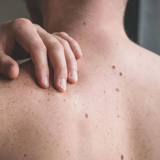
Seborrheic keratosis dermatitis is a common skin hyperplasia. It is often mistaken for a disease such as skin cancer because of its appearance that looks like warts, precancerous skin growths, or skin cancer. Dermoscopy of seborrheic keratosis dermatitis is crucial to identify seborrhei keratosis from other types of skin diseases. What is Seborrheic Keratosis Dermatitis?Seborrheic…
Dermoscopy of Seborrheic Keratosis vessels: early irritated Dermatitis - IBOOLO
Discover Dermoscopy of Seborrheic Keratosis vessels: early irritated Dermatitis in this concise guide. Learn how seborrheic keratosis dermoscopy identifies waxy surfaces and vascular patterns in early and irritated cases.
Dermoscopy of early Seborrheic Keratosis: Exploring Irritated and Early Seborrheic Keratosis
Dermoscopy of Seborrheic Keratosis: A Comprehensive Guide
Dermoscopy of seborrheic keratosis is a non-invasive diagnostic technique widely used to evaluate seborrheic keratosis. This article explores its core features, such as waxy surfaces and fingerprint-like structures, deeply analyzes the inflammatory manifestations of irritated seborrheic keratosis dermoscopy, and examines the early characteristics of early seborrheic keratosis dermoscopy. The article also provides operational guidelines and differential diagnostic techniques to help clinicians accurately identify lesions and provide safer, more efficient diagnostic support for patients.
What is Dermoscopy of Seborrheic Keratosis?
Dermoscopy of seborrheic keratosis is a non-invasive skin diagnostic technique specifically used to evaluate seborrheic keratosis (SK), a common benign skin tumor. The dermoscope magnifies the skin surface 10 to 100 times, helping clinicians observe the characteristic manifestations of seborrheic keratosis, such as waxy surfaces, fingerprint-like structures, and pigmentation. The primary clinical role of seborrheic keratosis dermoscopy is to distinguish it from malignant lesions (such as basal cell carcinoma or melanoma), avoiding unnecessary biopsies. This technique provides dermatologists with an efficient diagnostic tool, particularly suitable for identifying early or atypical seborrheic keratosis.
What Does the Equipment Structure and Workflow of Seborrheic Keratosis Dermoscopy Include?
Seborrheic keratosis dermoscopy relies on the structure and workflow of dermoscopic equipment to ensure accurate observation of lesion characteristics. The equipment typically includes the following parts:
Optical lens: Provides 10 to 100x magnification, displaying minute skin structures.
Light source system: Built-in LED lights or polarized light, reducing reflection and enhancing image contrast.
Image capture module: Some devices support high-resolution image capture for recording and analysis.
The workflow is as follows: The physician first cleans the lesion area, applies a coupling agent (such as alcohol or gel), then uses the dermoscope to observe the characteristics of seborrheic keratosis, such as waxy surfaces or fingerprint-like structures, and finally records images for subsequent analysis. This workflow for seborrheic keratosis dermoscopy ensures systematic and reliable diagnosis.
How Does the Working Principle of Seborrheic Keratosis Dermoscopy Operate?
The working principle of seborrheic keratosis dermoscopy is based on optical magnification and light optimization technology. The dermoscope displays details of the skin's surface and shallow layers through high magnification (typically 10 to 100x), while using polarized light or liquid media (such as gel) to reduce skin surface reflection, thereby improving image clarity. For seborrheic keratosis, dermoscopy can clearly present its characteristic appearances, such as waxy or "pasted-on" appearance, fingerprint-like structures, pigmentation, or corneal cysts. This technology enables doctors to more accurately identify typical features in seb keratosis dermoscopy, distinguishing it from malignant lesions and reducing the risk of misdiagnosis.
How to Use Dermoscopy to Diagnose Seborrheic Keratosis
Using dermoscopy to diagnose seborrheic keratosis requires following standardized operating procedures to ensure the accuracy of examination results. Here is a step-by-step guide:
1. Prepare the lesion area: Clean the lesion site with a mild cleanser to remove oils and dirt, ensuring the skin surface is clean.
2. Select the appropriate dermoscope: Choose a handheld or desktop dermoscopic device based on the size and location of the lesion.
3. Apply coupling agent: Apply alcohol or gel to the lesion area to reduce skin reflection and improve image clarity.
4. Observe characteristic manifestations: Carefully observe the characteristics of seborrheic keratosis through the dermoscope, such as waxy surfaces, fingerprint-like structures, or corneal cysts.
5. Record and analyze: Take dermoscopic images, record observations, and compare them with typical features of seborrheic keratosis.
Through these steps, seborrheic keratosis dermoscopy can provide reliable diagnostic evidence for clinicians.
What is the Application Range of Dermoscopy in Diagnosing Seborrheic Keratosis?
Dermoscopy has a wide range of applications in diagnosing seborrheic keratosis, primarily used to evaluate and differentiate seborrheic keratosis and its variant types. Seborrheic keratosis dermoscopy is applicable in the following scenarios:
Typical seborrheic keratosis: Identifying waxy surfaces, fingerprint-like structures, and corneal cysts.
Early seborrheic keratosis: Detecting subtle features in early lesions, such as slight pigmentation or surface roughness.
Irritated seborrheic keratosis: Observing erythema or erosion caused by inflammatory reactions, distinguishing it from malignant lesions.
Differentiation from malignant lesions: Distinguishing seborrheic keratosis from basal cell carcinoma or melanoma through characteristic manifestations.
Follow-up monitoring: Monitoring changes in seborrheic keratosis and evaluating whether further intervention is needed.
The non-invasive and high-resolution characteristics of seborrheic keratosis dermoscopy make it an important diagnostic tool for dermatologists.
Why are the Clinical Advantages of Seborrheic Keratosis Dermoscopy Prominent?
Seborrheic keratosis dermoscopy has significant advantages in clinical diagnosis. First, it is a non-invasive technique that allows observation of lesions without tissue removal, reducing patient discomfort and infection risk. Second, seborrheic keratosis dermoscopy can magnify lesion details, clearly presenting characteristic manifestations such as waxy surfaces and fingerprint-like structures, thereby improving diagnostic accuracy. Additionally, it helps differentiate it from malignant lesions (such as basal cell carcinoma or melanoma), reducing unnecessary biopsies. For early or irritated seborrheic keratosis, dermoscopic examination can identify lesions through subtle features, assisting doctors in formulating appropriate treatment plans. It is an important tool in dermatological diagnosis and treatment.
What Are the Typical Dermoscopic Features of Seborrheic Keratosis?
Seborrheic keratosis presents unique characteristic manifestations under dermoscopy, helping doctors make accurate diagnoses. The following are typical features in seborrheic keratosis dermoscopy:
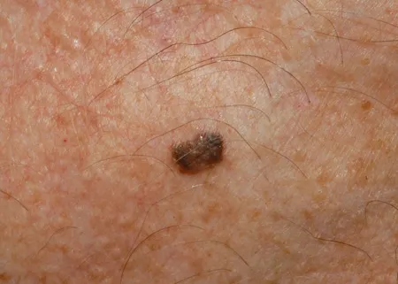
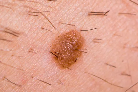
Waxy or "pasted-on" appearance: The lesion surface looks like it's "pasted" onto the skin, presenting a waxy luster.
Fingerprint-like structures: Linear or reticular structures similar to fingerprints, a signature presentation of seborrheic keratosis.
Corneal cysts: Small white or yellow keratin granules embedded in the lesion surface.
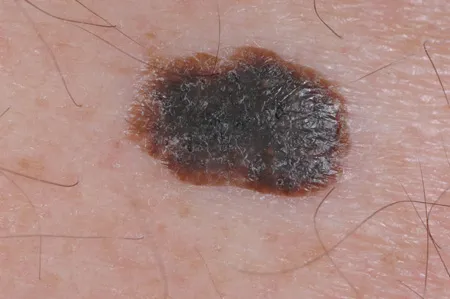
Pigmentation: Some lesions may have uniform brown or black pigmentation.
No obvious vessels: Unlike malignant lesions, seborrheic keratosis typically lacks irregular vascular patterns.
By identifying these features, seborrheic keratosis dermoscopy can effectively differentiate it from malignant lesions, reducing the risk of misdiagnosis.
What Are the Quality Control and Precautions for Seborrheic Keratosis Dermoscopy?
Quality control and operational precautions for seborrheic keratosis dermoscopy are crucial for ensuring diagnostic accuracy. The following are key considerations:
Clean the lesion area: Remove oils and dirt from the lesion site with a mild cleanser before examination to avoid affecting image clarity.
Select appropriate light source: Use polarized light mode to reduce reflection, ensuring waxy surfaces and fingerprint-like structures are clearly visible.
Avoid excessive pressure: Lightly touch the skin during examination, avoiding pressure on the lesion that could cause surface structure deformation.
Combine clinical information: Make judgments by integrating patient history and clinical manifestations, avoiding reliance solely on dermoscopic images.
Record complete data: Take and save high-quality images for follow-up and discussion with other doctors.
These standards can improve the reliability of seborrheic keratosis dermoscopy, providing solid support for diagnosis.
How to Maintain and Care for Dermoscopic Equipment
Maintenance and care of dermoscopic equipment are crucial for ensuring the accuracy of seborrheic keratosis dermoscopy and equipment longevity. Here is a step-by-step guide:
1. Clean the lens: After each use, gently wipe the lens with a dedicated lens cleaning cloth and solution to remove dust and fingerprints.
2. Check the light source: Confirm that the dermoscope's light source has normal brightness; replace bulbs or batteries promptly if the light dims.
3. Avoid physical damage: Place the dermoscope in its protective case after use to avoid drops or collisions that could damage internal components.
4. Maintain dry storage: Store the device in a dry, light-protected environment, avoiding moisture or high temperatures that could affect performance.
5. Regular calibration: Send to a professional institution for calibration every 6-12 months to ensure magnification and imaging accuracy.
Through these steps, seborrheic keratosis dermoscopy equipment can maintain optimal condition, providing reliable support for diagnosis.
What Are the Dermoscopic Manifestations of Inflammatory Seborrheic Keratosis?
Irritated seborrheic keratosis dermoscopy presents different manifestations from typical seborrheic keratosis under dermoscopy, requiring special attention to avoid misdiagnosis. In seborrheic keratosis dermoscopy, the characteristics of inflammatory lesions include:
Erythema and inflammation: Erythema may appear around the lesion, accompanied by mild swelling.
Surface erosion or crusting: Inflammation causes surface damage, possibly with erosion or crust formation.
Slight vascular proliferation: Unlike typical seborrheic keratosis, inflammatory lesions may present with dotted or short linear vessels.
Uneven pigmentation: Inflammation may intensify pigmentation, presenting uneven brown or black coloration.
These manifestations make irritated seborrheic keratosis dermoscopy more challenging, requiring physicians to carefully differentiate in conjunction with clinical history to rule out the possibility of malignant lesions.
What Are the Key Points in Dermoscopic Diagnosis of Early Seborrheic Keratosis?
Early seborrheic keratosis dermoscopy is crucial for timely identification of lesions. The diagnostic points of early seborrheic keratosis dermoscopy include the following features:
Mild surface roughness: The lesion surface may present subtle granular or waxy texture.
Uniform pigmentation: Early lesions typically have mild brown or black pigmentation, distributed relatively uniformly.
Emerging fingerprint-like structures: Indistinct fingerprint-like or reticular structures may be observed, suggesting lesion characteristics.
No obvious vessels: Early seborrheic keratosis typically lacks significant vascular patterns, contrasting with malignant lesions.
By identifying these subtle features, early seborrheic keratosis dermoscopy can help doctors make accurate diagnoses in the initial stages of lesions, avoiding misdiagnosis as malignant lesions.
What is the Analysis of Vascular Features in Seborrheic Keratosis Dermoscopy?
The analysis of vascular features in seborrheic keratosis dermoscopy is an important part of differential diagnosis. Unlike malignant lesions, seborrheic keratosis typically lacks significant vascular patterns, but subtle changes may appear in certain conditions. The following are the vascular features in seborrheic keratosis dermoscopy:
Sparse vessels: Typical seborrheic keratosis usually has no obvious vessels or only a few dotted vessels.
Vessels in irritated lesions: In irritated seborrheic keratosis, short linear or dotted vessels may appear, accompanied by inflammatory reactions.
No irregular patterns: Unlike the polymorphic vessels in melanoma, the vascular pattern in seborrheic keratosis is relatively simple.
By analyzing these vascular features, seborrheic keratosis dermoscopy vessels can help doctors distinguish it from malignant lesions, reducing the risk of misdiagnosis.
How to Perform Dermoscopic Differential Diagnosis Between Seborrheic Keratosis and Melanoma
Dermoscopic differential diagnosis between seborrheic keratosis and melanoma is an important application of seborrheic keratosis dermoscopy. The following are key differential points:
Surface structure: Seborrheic keratosis presents waxy or fingerprint-like structures, while melanoma often has irregular elevations or surface defects.
Pigment distribution: Seborrheic keratosis has uniform pigmentation, whereas melanoma presents as irregular pigment networks and multiple colors (brown, black, red).
Vascular pattern: Seborrheic keratosis has sparse or no obvious vessels, while melanoma often has irregular polymorphic vessels.
Border features: Seborrheic keratosis has clear borders, while melanoma has blurred and asymmetric borders.
Through these features, seborrheic keratosis dermoscopy can help doctors accurately distinguish between the two lesions, avoiding unnecessary invasive examinations.
What is the Comparison of Dermoscopic Presentations Among Different Types of Seborrheic Keratosis?
Different types of seborrheic keratosis present diverse manifestations under dermoscopy, understanding these differences aids in precise diagnosis. The following is a comparison of types in seborrheic keratosis dermoscopy:
Typical seborrheic keratosis: Waxy surface, fingerprint-like structures, corneal cysts, uniform pigmentation.
Irritated seborrheic keratosis: Erythema, erosion, or crusting, possibly accompanied by short linear vessels.
Early seborrheic keratosis: Slightly rough surface, indistinct pigmentation, emerging fingerprint-like structures.
Pigmented seborrheic keratosis: Deep brown or black pigmentation, possibly with reticular structures on the surface.
By comparing these presentations, seborrheic keratosis dermoscopy can help doctors identify the lesion type and formulate targeted management plans.
How is Dermoscopy Applied in Seborrheic Keratosis Treatment Follow-up?
Dermoscopy plays an important role in seborrheic keratosis treatment follow-up, helping doctors evaluate treatment effects and monitor lesion changes. For example, after treating seborrheic keratosis with cryotherapy or laser therapy, dermoscopy can observe whether the lesion has subsided or if there are signs of recurrence. The specific applications of seborrheic keratosis dermoscopy include:
Monitoring surface changes: Observing whether the waxy surface or fingerprint-like structures have disappeared.
Evaluating inflammatory responses: Checking if erythema or erosion in irritated lesions has improved.
Identifying residual lesions: Determining whether further treatment is needed through subtle features.
Recording dynamic changes: Taking dermoscopic images to compare lesion features before and after treatment.
This non-invasive technique provides reliable support for the long-term management of seborrheic keratosis.
What is the Analysis of Dermoscopic Features in Inflammatory Seborrheic Keratosis?
Irritated seborrheic keratosis dermoscopy presents different features from typical seborrheic keratosis under dermoscopy, requiring careful analysis to ensure accurate diagnosis. The features of irritated seborrheic keratosis dermoscopy include:
Erythema and inflammatory reaction: Obvious erythema around the lesion, accompanied by mild swelling.
Surface erosion or crusting: Inflammation causes surface damage, possibly presenting erosion, exudation, or crusting.
Uneven pigmentation: Inflammation may intensify pigmentation, presenting uneven brown or black coloration.
Slight vascular proliferation: Dotted or short linear vessels may be observed, differing from the non-vascular features of typical seborrheic keratosis.
By identifying these features, irritated seborrheic keratosis dermoscopy can help doctors accurately assess the lesion status.
How to Perform Dermoscopic Differential Diagnosis of Inflammatory Seborrheic Keratosis?
Dermoscopic differential diagnosis of inflammatory seborrheic keratosis is an important clinical step, as its features may resemble malignant lesions (such as basal cell carcinoma). The following are key differential points:
Surface features: Inflammatory seborrheic keratosis often has erosion or crusting, while basal cell carcinoma may present superficial ulcers and leaf-like areas.
Vascular pattern: Inflammatory seborrheic keratosis may have short linear vessels, while basal cell carcinoma primarily has arborizing vessels.
Pigment distribution: The pigmentation in inflammatory seborrheic keratosis is uneven but still relatively uniform, while malignant lesions like melanoma have irregular pigment networks.
Borders: Inflammatory seborrheic keratosis has relatively clear borders, while malignant lesions typically have blurred borders.
Through these features, irritated seborrheic keratosis dermoscopy can help doctors accurately differentiate lesion types, avoiding misdiagnosis.
How is Dynamic Observation in Irritated Seborrheic Keratosis Dermoscopy Performed?
Dynamic observation in irritated seborrheic keratosis dermoscopy is crucial for evaluating lesion progression and treatment effects. Irritated seborrheic keratosis dermoscopy can monitor lesion changes through regular follow-ups, for example:
Inflammation subsidence: Observing whether erythema and swelling have reduced, and if surface erosion has healed.
Vascular changes: Monitoring whether short linear or dotted vessels have decreased, indicating inflammation relief.
Pigment evolution: Recording whether pigmentation tends to become uniform, judging lesion stability.
Structure restoration: Checking whether typical seborrheic keratosis features like waxy surfaces or fingerprint-like structures have been restored.
Through dynamic observation, irritated seborrheic keratosis dermoscopy can help doctors assess whether further intervention is needed, optimizing treatment plans.
What Are the Vascular Patterns in Irritated Seborrheic Keratosis Dermoscopy?
Analysis of vascular patterns in irritated seborrheic keratosis dermoscopy is an important basis for differential diagnosis. Unlike typical seborrheic keratosis, inflammatory lesions may present vascular changes due to inflammatory reactions. The following are the characteristics of its vascular patterns:
Dotted vessels: Small dotted vessels may appear in inflamed areas, distributed unevenly.
Short linear vessels: Some lesions may show short and straight linear vessels, accompanied by erythema.
No arborizing vessels: Unlike the arborizing vessels in basal cell carcinoma, the vascular pattern in inflammatory seborrheic keratosis is relatively simple.
Inflammation-related: Vascular proliferation is usually related to the degree of inflammation and may decrease after inflammation subsides.
These vascular features allow seborrheic keratosis dermoscopy vessels to more accurately assess the nature of lesions.
How Can Dermoscopic Diagnostic Skills for Difficult Cases Be Enhanced?
Seborrheic keratosis dermoscopy for difficult cases requires special skills to improve diagnostic accuracy. Here are some practical tips:
Multi-angle observation: Observe lesions from different angles to ensure no subtle features are missed, such as fingerprint-like structures or slight vessels.
Integrate clinical information: Make judgments by integrating patient history (such as lesion evolution time, irritating factors) and clinical manifestations.
Use polarized light: Switch to polarized light mode to reduce reflection and more clearly observe pigmentation and vascular patterns.
Compare with typical features: Compare difficult cases with typical seborrheic keratosis features (such as waxy surfaces) to find differences.
Record dynamic changes: Take images at multiple time points to observe lesion changes over time, aiding diagnosis.
Through these skills, seborrheic keratosis dermoscopy can more effectively address difficult cases, reducing the risk of misdiagnosis.
This article comprehensively analyzes the application of dermoscopy of seborrheic keratosis in diagnosis, covering its typical features, inflammatory manifestations in irritated seborrheic keratosis dermoscopy, and early features in early seborrheic keratosis dermoscopy. Through operational standards, differential diagnostic points, and dynamic observation methods, the article provides practical guidance for medical professionals, helping to improve diagnostic accuracy and treatment follow-up efficiency, emphasizing the important value of dermoscopic technology in distinguishing between benign and malignant lesions.
Recommended reading
amelanotic melanoma dermoscopy – IBOOLO
IBOOLO is a camera lens manufacturer based in China with more than 11+ years of experience in manufacturing, catering to a variety of requirements. We have become experts in the design and manufacture of a wide variety of Dermatoscope, Microscope, Macro lens and Woods Lamp.
China's Premier Smartphone Dermoscopy Basal Cell Carcinoma Manufacturing Products Supply - IBOOLO
Through masterful production techniques, our China products supply delivers advanced yet smartphone dermoscopy basal cell carcinoma merging cutting-edge optics with compact, ergonomic frames ideal for transport and mobility.
China Skin Cancer Dermoscopy Products Supply Specializes in Professional Items - IBOOLO
Our China products supply creates clinical quality Professional skin cancer dermoscopys enabling powerful skin magnification from anywhere through thoughtful craftsmanship.
Seborrheic keratosis dermatitis is a common skin hyperplasia. It is often mistaken for a disease such as skin cancer because of its appearance that looks like warts, precancerous skin growths, or skin cancer. Dermoscopy of seborrheic keratosis dermatitis is crucial to identify seborrhei keratosis from other types of skin diseases.
What is Seborrheic Keratosis Dermatitis?
Seborrheic keratosis dermatitis is a kind of benign epidermal hyperplasia caused by keratinocyte hyperplasia. Seborrheic keratosis dermatitis is a type of non-cancerous benign of skin disease. Seborrheic keratosis dermatitis is harmless.
Seborrheic keratosis dermatitis is known as senile warts, senile spots, also known as basal cell papilloma. Because it mainly occurs in adults over the age of 40, it often appears as people grow older.
What are the Clinical Feature of Seborrheic Keratosis Dermatitis?
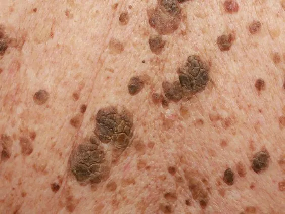
Seborrheic keratosis dermatitis is painless and it usually appears brown, black, or light tan. Its growth appears waxy or scaly and is slightly raised. They can gradually appear on various parts of the body, mostly on the face, neck, chest, or back.
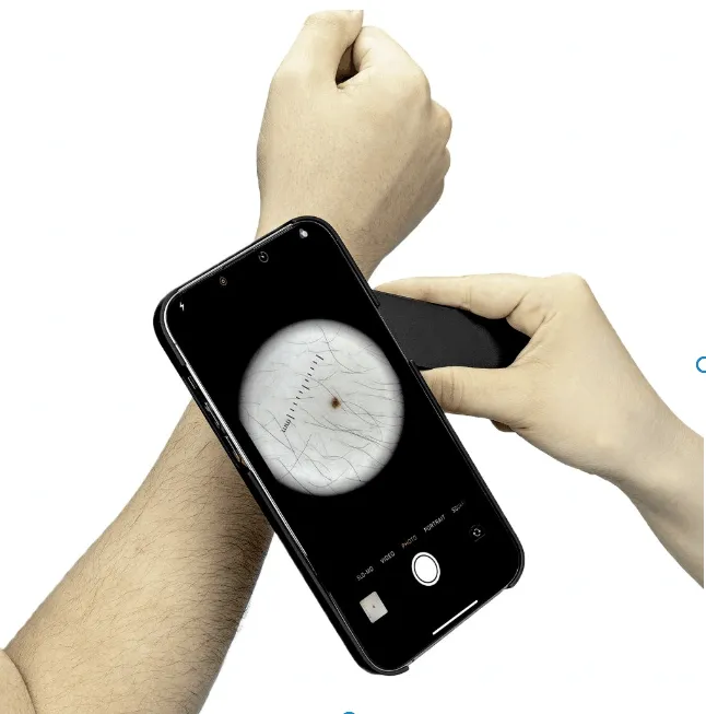
Why is It Necessary to Use a Dermoscopy of Seborrheic Keratosis Dermatitis?
Dermatoscope is a non-invasive technique that allows dermatologists to closely examine seborrheic keratosis dermatitis more accurately and precisely. Especially dermoscopy of seborrheic keratosis dermatitis greatly enhance the vision of some locations that hard-to-reach by naked eyes, such as details in lesions. Dermatoscope magnifys and brightens shapes and structures of lesions. Dermoscopy of seborrheic keratosis dermatitis increases the confidence of doctors and patients about the skin disease and avoids unnecessary anxiety and treatment. So it is really necessary to use a dermoscopy of seborrheic keratosis.
Typical Features of Dermoscopy of Seborrheic Keratosis Dermatitis
dermoscopy plays a crucial role in identifying seborrheic keratosis dermatitis. There are some typical features of dermoscopy of seborrheic keratosis dermatitis as below:
Special pattern: Typical “gyrigrain” or “fat-like” pattern.
Hair follicle openings: Visible hair follicle openings.
Structure: Edge ring structure, Light brown fingerprint-like parallel structures.
Prominent blood vessels: In some forms of seborrheic keratosis dermatitis, a halo of lobules surrounds tiny, hairpin-like capillaries.
Miliary cysts: These cysts may appear as small white stars or larger, yellowish turbidity.
Other features like: cracks/ridges, blue-gray balls, irregular crypts, weak or pseudo-network.
Dermoscopy of seborrheic keratosis dermatitis is very helpful and reliable for distinguish seborrheic keratosis dermatitis from other skin diseases.
How to differentiate between seborrheic keratosis dermatitis and melanoma?
Seborrheic kearatosis dermatitis will not transfer into melanoma. But both of seborrheic keratosis dermatitis and melanoma can be brown or black color, so the two can be easily be mistake from each other.
There are some important differences between seborrheic keratosis dermatitis and melanoma, from their numbers, appearances, locations causes, etc.
Comparison the apearances of seborrheic keratosis dermatitis and melanoma
Numbers: Seborrheic keratoses dermatitis: Seborrheic keratoses dermatitis often appear in numbers of two or more
Melanoma: Melanoma is usually appear in single.
Shapes: Seborrheic keratoses dermatitis: Seborrheic keratoses dermatitis usually shows round or oval shaped.
Melanoma: Irregular shape, asymmetry in shape is the typical features of melanoma.
Colors: Seborrheic keratoses dermatitis: Seborrheic keratoses dermatitis colors in light tan, brown, or black.
Melanoma: Melanoma is commonly display multiple colors like pink, red, white, blue or mixed color within the same one.
Size: Seborrheic keratosis dermatitis: Seborrheic keratosis dermatitis varies in size from very small to big, and its size will not changed as time goes on.
Melanoma: Melanoma is in bigger size than 1/4 inch, and its size will grow over time.
Surface: Seborrheic keratosis dermatitis: Seborrheic keratosis dermatitis has waxy or scaly surface, slightly elevated above the skin surface.
Melanoma: Melanoma tends to be smooth with blurred, ragged border.
Pain: Seborrheic keratosis dermatitis:Seborrheic keratosis dermatitis is painless
Melanoma: Some of melanoma feel hurt, while some of melanoma feel no any pain or discomfort.
Evolving: Seborrheic keratosis dermatitis:Seborrheic keratosis dermatitis maintains the same always.
Melanoma: Melanoma may looks different from its beginning, and it may change in its shape, size or color.
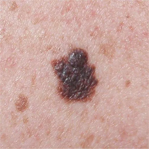
Comparison the locations of seborrheic keratosis dermatitis and melanoma
Seborrheic keratosis dermatitis: Seborrheic keratosis dermatitis mostly displays on the face, neck, chest, or back.
Melanoma: Melanoma Melanoma can appear in anywhere on the skin,mostly on chest, black, legs, arms, face, necks, and even eyes.
Comparison the causes of seborrheic keratosis dermatitis and melanoma
Causes: The primary risk factor for seborrheic keratoses is age. Other risk factors include: sunburn, skin irritation and friction, pregnancy, hormone therapy, some medications, genetic mutation, a family history of seborrheic keratosis
How is seborrheic keratosis dermatitis diagnosed?
To diagnose seborrheic keratosis dermatitis, skin doctors will get information from your family history of skin disease and take observation of it through a vision aiding tool called dermatoscope. Dermatoscope is a small handheld lighted medical microscope that allows a more precise and deeper view of skin diseases by high magnification and a powerful glare-free lighting system. If it is necessary, a biopsy will be needed for seborrheic keratosis.
People usually also take dermoscopy of seborrheic keratosis dermatitis for self-examination on skin. Any unusual findings or changes occur, have dermatologist checked for a further evaluation. Dermoscopy of seborrheic keratosis dermatitis plays a significant role in physical exam.
Application of dermoscopy of seborrheic keratosis dermatitis
A dermoscope is a main device used to examine skin diseases, like seborrheic keratosis dermatitis. In the diagnosis and treatment of seborrheic keratosis dermatitis, dermoscopy of seborrheic keratosis dermatitis is widely used in the following aspects:
Monitoring: For patients who have already been diagnosed with seborrheic keratosis dermatitis, dermoscopy can be used to monitor the whole process as time goes on. If any changes happen, a further step should be taken.
Feedback: Skin doctor can compare images from dermoscopy of seborrheic keratosis dermatitis over different times to assess the effectiveness of the treatment of seborrheic keratosis dermatitis and then decide if the treatment needs to be adjusted or not.
Treatment aid: When treating seborrheic keratosis dermatitis, images can be clearly and precisely showed by dermoscopy of seborrheic keratosis dermatitis. It greatly enhanced the patience of skin doctors and patients.
Seborrheic keratosis dermatitis is a harmless skin disease which will not cause skin cancer. But skin doctors should have it accurately diagnosed by dermoscope. Dermoscopy of seborrheic keratosis dermatitis is very important to differentiate seborrheic keratosis dermatitis from other skin diseases. Hence, skin doctors can remove seborrheic keratoss surely and safely for some aesthetic reasons.
It is vital to develop the habit of use of dermoscopy of seborrheic keratosis dermatitis. In addition, paying more attention to a regular skin examination is also necessary in our daily life. People should remain vigilant at all time to keep the health of the skin.


