


Article
Dermoscopy of Negative Network
The negative network is represented by serpiginous, interconnecting, broadened hypopigmented lines around elongated and curvilinear globules. In a dermatoscope, a positive usually indicates that certain abnormal pathological features are detected. The benefit of the diagnosis of skin lesions is that a negative finding can help rule out melanoma, basal cell carcinoma or squamous cell carcinoma….
The negative network is represented by serpiginous, interconnecting, broadened hypopigmented lines around elongated and curvilinear globules. In a dermatoscope, a positive usually indicates that certain abnormal pathological features are detected. The benefit of the diagnosis of skin lesions is that a negative finding can help rule out melanoma, basal cell carcinoma or squamous cell carcinoma.
Fundamentals of Dermoscopy
The basic principle of dermoscopy is transillumination of a lesion in order to study it with high magnification to visualize subtle features. Light incident on a surface like the skin may be reflected, refracted, diffracted and/or absorbed. The physical properties of the skin influence these phenomena. Most light incident on dry, scaly skin is reflected, but smooth, oily skin allows light to pass through to reach the deeper dermis. Application of a linkage or immersion fluid over the skin, enhances translucency and improves visibility of subsurface skin structures of the lesion under investigation.

Dermoscopic Features of Negative Network
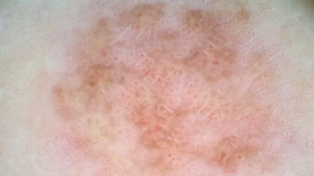
The “negative” of the pigmented network (also known as reverse or inverse network) consists of relatively lighter areas comprising the apparent grid of the network and relatively darker areas filling the apparent “holes”. The lighter grid lines tend to be serpiginous and the darker areas, when viewed in isolation, resemble elongated tubular or curved globules. Histopathologically, the negative network appears to correspond to thin elongated rete ridges accompanied by large melanocytic nests within a widened papillary dermis or to bridging of rete ridges. The negative network is highly specific for melanoma (95% specific), especially for a melanoma arising in a nevus.
Clinical Significance of the Negative Network
The dermoscopic descriptor “negative pigment network” (NPN) has been reported in several types of melanocytic and non-melanocytic lesions, although it has a higher frequency of association with melanoma and Spitz naevus. In a study of 401 consecutive melanomas, excluding facial, acral and mucosal locations, the frequency and variability of NPN were investigated, and the results of NPN correlated with clinical and histopathological data. NPN of any extension was found in 27% of melanomas, most frequently invasive and arising from a naevus on the trunk of young subjects. Seven percent of melanomas in the study population showed presence of NPN in more than half of the lesion area; most of these did not show typical dermoscopic melanoma features.
Steps in Performing Dermoscopy
When skin magnification is performed, the magnification of the dermatoscope is adjusted according to the size of the examined area and the details required. Observe the window of the dermatoscope until the area under observation is clear. As well as to avoid too close contact between the dermatoscope and the skin, so as not to affect the observation effect or produce errors.
Although the non-invasive examination of dermoscopy can provide a lot of information, the final diagnosis may still need to be confirmed by skin biopsy and pathology. For suspicious lesions found during dermoscopy, doctors may consider further pathological section examination.
How to Analyse Dermatological Images to Identify Negative Network
The Negative network is defined by serpiginous lighter grid lines that are linked with hyperpigmented, elongated to curvilinear globules. The negative network from a histological point of view seems to match with the thin elongated and hypopigmented rete ridges that are bridging and encircling large melanocytic nests in a widened dermal papillae. There are Dermoscopy tumour simulacra and mimics. False positive diagnosis may lead to unnecessary excisions. Missing a cancer case is much more risky as it may lead to severe consequences for the patient and the doctor as well as false negative diagnosis.

Negative Results and Other Diagnostic Methods
Dermoscopy is widely used for the evaluation of melanotic lesions and also as an aid in the diagnosis of vascular diseases and parasitic skin infections. It has the advantage of being non-invasive and rapid, but has limited diagnostic value for non-pigmented lesions. The technique of Histopathological examination secures tissue samples by skin biopsy for pathological examination. Even though it is an invasive technique it is the gold standard for the diagnosis of many difficult skin diseases like skin cancer, lupus erythematosus, pemphigus; however, it is an invasive process and can be prone to sampling bias. Only in the case of a high clinical suspicion of a disease dermoscopy can be negative and further skin biopsy, PCR or immunological testing can be done. If direct microscopy is negative in a dermatomycosis case but there is high suspicion of the disease, fungal culture may be done.
Negative Networks in Skin Health Monitoring
A baseline of an individual’s health is established through skin health surveillance. The result that does not show a suspicious melanotic lesion is negative and can be used as a reference for future surveillance. A number of patients are concerned with the outcome of skin tests, and doctors should tell them that a negative result indicates that there is no evidence of disease at this time, not no disease at all time. It is best to have the patients do check up regularly dermoscopy and have the patients save dermoscopic images as recommended by their doctors. It can then be compared with the digital images of different periods of time in order to detect the lesions.

Challenges and Misconceptions of Negative Results
Some analyses are not a magic wand; each one is different and has its own sensitivity and specificity. It is also important to understand that it is impossible to exclude all diseases with 100% confidence from a result. For example, not all melanomas are detectable in the early or subclinical stages, so dermoscopy may not detect the lesion. Dermoscopy can be used in conjunction with histological biopsy, or PCR with fungal culture, which can increase the diagnostic yield and prevent wrong diagnosis with negative findings.
Conclusion
Dermoscopy results negative may provide a less anxious patient with skin cancer or other serious skin diseases. High risk group or family history of skin diseases patients should also maintain routine check up schedule with the doctor even if the patient has no lesions. There is no zero risk for the condition from a negative result, and this is particularly so for those with high prevalence risk factors for skin diseases. Hence, patients should still practice good skin health through education, lifestyle changes.
Negative Network Dermoscopy: A Guide to Diagnosis, Pitfalls, and Advanced Techniques
Negative network dermoscopy is a critical tool for dermatologists, offering insights into pigmented lesions that may indicate melanoma or other skin conditions. This pattern, characterized by the absence or reversal of typical pigment networks, can be challenging to interpret but is essential for accurate diagnosis. This guide explores the clinical applications of negative network dermoscopy, from early melanoma detection to differentiating melanocytic from non-melanocytic lesions. Learn about proper techniques, common pitfalls, and advanced imaging methods like OCT and confocal microscopy. Whether you're a clinician or a patient, discover how negative network dermoscopy enhances diagnostic precision and improves outcomes.
What is Negative Network in Dermoscopy: A Comprehensive Guide for Dermatologists
In dermoscopy, the negative network is a significant visual pattern. But what exactly is it? A negative network refers to a pattern where the pigmented lines and dots that typically form a network-like structure in normal skin or certain benign lesions are absent or appear as a reverse image. For example, in a typical pigmented nevus, there is often a well-defined network of pigmented lines. However, in cases where a negative network is present, these lines are either not there or are seen as lighter areas against a darker background.
Why is understanding the negative network crucial for dermatologists? It can be an important diagnostic clue. In some cases, a negative network may be associated with specific skin conditions, such as certain types of melanomas. By recognizing this pattern, dermatologists can make more informed decisions about further testing or treatment. It helps in differentiating between benign and malignant skin lesions, which is essential for providing the best possible patient care.
What is the Structure of Dermoscopes Optimal for Negative Network Visualization?
The structure of a dermoscope plays a vital role in visualizing the negative network. First, a high-quality lens is essential. Why is the lens so important? A good lens provides clear and sharp images, allowing dermatologists to accurately identify the subtle patterns of the negative network. Look for dermoscopes with lenses that have a high numerical aperture, as this enables better resolution and contrast.
Illumination is another key aspect. How does illumination affect negative network visualization? Dermoscopes with proper illumination, such as polarized light sources, can enhance the visibility of the negative network. Polarized light reduces surface reflections, making it easier to see the underlying skin structures. Some dermoscopes also offer adjustable illumination intensity, which can be customized depending on the patient's skin type and the specific lesion being examined.
The magnification power of the dermoscope is also crucial. What magnification is ideal? For visualizing the negative network, a magnification range of 10 - 40x is often recommended. This range allows for a detailed view of the skin's surface and subsurface structures, where the negative network patterns are most prominent.
How to Follow a Clinical Workflow for Identifying Negative Network Patterns in Pigmented Lesions
When dealing with pigmented lesions and aiming to identify negative network patterns, a well-defined clinical workflow is essential. Step by step:
Initial Patient Assessment:
First, obtain a detailed patient history. Why is this important? Information about the patient's age, sun exposure history, family history of skin cancer, and any changes in the pigmented lesion over time can provide valuable context. For example, a sudden change in a mole's appearance in a patient with a high sun exposure history may increase the suspicion of malignancy.
Gross Visual Inspection:
Before using the dermoscope, visually examine the pigmented lesion. Look for characteristics such as size, shape, colour, and border irregularity. This initial inspection can give an idea of whether further investigation with dermoscopy for negative network patterns is warranted.
Dermoscopic Examination:
Gently place the dermoscope on the skin surface over the pigmented lesion. Adjust the focus and illumination settings according to the guidelines for optimal negative network visualization. Look carefully for the absence or reverse pattern of the typical pigmented network. If a negative network is detected, note its location, extent, and any associated features within the lesion.
How Do Dermoscopy Working Principles Reveal Negative Network Structures?
Dermoscopy works on several principles to reveal negative network structures. One of the main principles is the use of polarized or non-polarized light. How does light help? When light is shone on the skin through the dermoscope, it interacts with the skin's layers. In normal skin, the light is absorbed, scattered, and reflected in a way that creates the appearance of a pigmented network. However, in areas where a negative network is present, the light behaves differently.
For example, in non-polarized light dermoscopy, the negative network may appear as areas where less light is absorbed by the pigmented structures, resulting in lighter-coloured regions. In polarized light dermoscopy, the reduction of surface reflections allows for a better view of the underlying structures. The negative network may be more clearly visible in areas with a different light-scattering pattern compared to the surrounding skin.
Another principle is magnification. Why is magnification important? By magnifying the skin surface, dermoscopy reveals the fine details of the skin's pigmented structures. This magnification enables dermatologists to identify the absence or abnormal arrangement of the pigmented network that characterizes the negative network.
What is the Proper Technique for Visualizing Negative Network Patterns During Dermoscopic Examination?
To properly visualize negative network patterns during a dermoscopic examination, several techniques should be followed. First, ensure a proper skin-dermoscope contact. How do you achieve this? Gently place the dermoscope on the skin surface without applying too much pressure. Applying excessive pressure can distort the skin and affect the appearance of the negative network.
Adjust the focus carefully. Why is focusing important? A clear focus is essential to accurately identify the subtle patterns of the negative network. Start with a low magnification and gradually increase it while adjusting the focus until the skin structures are visible.
Pay attention to the illumination settings. What should you do with illumination? As mentioned earlier, for better visualization of the negative network, use the appropriate type of light (polarized or non-polarized) and adjust the intensity. If the light is too bright or too dim, it can make it difficult to see the negative network.
Finally, scan the entire pigmented lesion systematically. In a list format:
Start from one edge of the lesion.
Move the dermoscope slowly across the lesion, ensuring that every part of the lesion is examined.
Look for any areas that show the characteristic features of a negative network, such as the absence or reverse pattern of the pigmented network.
What Are the Clinical Applications of Negative Network Detection in Melanocytic Lesions?
In the realm of melanocytic lesions, the detection of negative network patterns has several crucial clinical applications. One significant application is in the early identification of melanoma risk. How does this work? Melanomas often present with abnormal pigmentation patterns, and the negative network can be an early sign. For example, in a suspicious mole, the presence of a negative network might prompt further diagnostic tests such as a biopsy. This early detection can significantly improve the prognosis for patients, as melanoma is more treatable in its early stages.
Negative network detection also aids in monitoring the progression of melanocytic lesions over time. Why is this important? By regularly examining lesions for changes in the negative network pattern, dermatologists can assess whether a lesion is stable, improving, or worsening. In cases where a negative network becomes more prominent or spreads within a lesion, it could indicate a more aggressive form of the melanocytic condition, guiding decisions on treatment intensity and frequency.
How Accurate Is the Negative Network in Differentiating Melanoma from Benign Lesions?
The negative network is considered a valuable tool in differentiating melanoma from benign lesions, but its accuracy is influenced by various factors. In terms of sensitivity, studies have shown that the presence of a negative network can be associated with melanoma in a significant number of cases. How does this translate clinically? When dermatologists observe a negative network in a pigmented lesion, there is a higher likelihood that the lesion may be malignant. However, it's not a foolproof indicator. Some benign lesions may also exhibit features that resemble a negative network, reducing the overall specificity.
To improve diagnostic accuracy, dermatologists often combine the observation of the negative network with other dermoscopic features. What are these features? This includes looking at asymmetry, border irregularity, colour variegation, and the presence of other abnormal structures within the lesion. By considering multiple factors, the accuracy of differentiating melanoma from benign lesions can be enhanced, leading to more reliable diagnoses and better patient care.
What Are the Key Features to Look for in Dermoscopes for Optimal Negative Network Visualization?
When seeking a dermoscope for optimal negative network visualization, several key features should be considered. A high-quality optical system is fundamental. Why is this so important? A top-notch optical system, including lenses with excellent resolution and contrast capabilities, allows for clear visualization of the fine details of the negative network. Look for dermoscopes with lenses that can minimize chromatic aberration, ensuring that colours are accurately represented and the negative network patterns are distinct.
Illumination features play a critical role as well. How does illumination affect negative network visualization? Dermoscopes with variable illumination options, such as switchable polarized and non-polarized light, are highly beneficial. Polarized light reduces surface glare, making it easier to see the underlying negative network patterns. Additionally, adjustable illumination intensity enables customization based on the patient's skin type and the specific characteristics of the lesion. For example, darker skin types may require higher illumination intensity to effectively visualize the negative network.
Magnification flexibility is another important aspect. What magnification range is ideal? A dermoscope that offers a magnification range from around 10x to 40x is often preferred. Lower magnifications can be used for a broad overview of the lesion, while higher magnifications allow for a detailed examination of the negative network structures, revealing subtle differences that may be crucial for diagnosis.
What Are the Quality Standards in Dermoscopic Evaluation of Negative Network Patterns?
Quality standards in dermoscopic evaluation of negative network patterns are essential for reliable diagnoses. Standardized image acquisition is a starting point. How should images be acquired? Dermoscopists should follow a set protocol for placing the dermoscope on the skin, ensuring consistent pressure and contact. This helps in obtaining reproducible images, which are crucial for accurate comparison over time. The images should also be of high resolution, with proper focus and exposure, to display the negative network patterns.
Accurate interpretation of negative network patterns is another key quality standard. Dermoscopists need to be well-trained in recognizing the nuances of the negative network. In a list format:
They should be able to distinguish between true negative network patterns and artefacts.
Understanding the association between the negative network and different skin conditions, such as melanoma and certain benign melanocytic lesions, is vital.
Continuing education and participation in quality assurance programs can help dermoscopists stay updated on the latest knowledge regarding negative network evaluation.
Documentation of the dermoscopic findings related to the negative network also adheres to quality standards. All relevant information, including the location of the lesion, the characteristics of the negative network (such as its extent and distribution), and any associated features, should be recorded. This documentation serves as a reference for future consultations and for tracking the progress of the patient's condition.
What Precautions Should Be Taken When Interpreting Negative Network Patterns Across Different Skin Types?
Interpretation of negative network patterns requires special precautions when dealing with different skin types. In darker skin types, the natural pigmentation can sometimes mask or mimic the appearance of a negative network. How can this be addressed? Dermatologists need to be aware of the normal variations in skin pigmentation for different ethnic groups. For example, in individuals with darker skin, there may be more prominent pigmentation around hair follicles, which could be mistaken for a negative network. Using appropriate illumination settings, such as increasing the intensity of polarized light, can help in differentiating between normal pigmentation and true negative network patterns.
In lighter skin types, on the other hand, the negative network may be more difficult to detect due to the lower contrast. What should be done? Dermoscopists may need to adjust the magnification and focus more precisely to identify the subtle negative network patterns. Additionally, using image enhancement techniques, if available on the dermoscope, can improve the visibility of the negative network in lighter-skinned patients. It's also important to consider the patient's overall skin condition, such as the presence of sun damage or other skin disorders, as these can affect the appearance of the negative network and its interpretation.
How to Maintain Dermoscopic Equipment for Precise Negative Network Visualization?
Maintaining dermoscopic equipment is crucial for consistently accurate visualization of negative network patterns. Regular cleaning is the first line of maintenance. Step by step, start by powering off the device. Use a soft, lint-free cloth slightly dampened with isopropyl alcohol to gently wipe the lens. Why is lens cleaning important? A dirty lens can distort the image, making it difficult to see the negative network. After cleaning the lens, wipe down the body of the dermoscope to remove any fingerprints or debris.
Calibration is another essential aspect. How often should calibration be done? Depending on the manufacturer's recommendations, it is typically advisable to calibrate the dermoscope at least once a year or after a certain number of uses. Calibration ensures that the magnification and colour representation are accurate. This is vital because incorrect magnification can make the negative network appear larger or smaller than it is, and inaccurate colour representation may lead to misinterpretation of the pattern.
Inspecting for physical damage is also necessary. What should you look for? Check for any cracks in the lens, loose parts, or damage to the light source. Even a small crack in the lens can significantly affect image quality. If any damage is detected, the equipment should be sent for repair immediately to avoid compromised negative network visualization.
Can Digital Dermoscopy Track Changes in Negative Network Patterns Over Time?
Digital dermoscopy offers great potential for tracking changes in negative network patterns over time. How does it work? Digital dermoscopes can capture high - resolution images of skin lesions with negative network patterns. These images can be stored in a patient's digital file. By comparing images taken at different time points, dermatologists can observe any alterations in the negative network. For example, if the negative network becomes more widespread or if the thickness of the lines within the negative network changes, it could indicate a progression of the underlying skin condition.
The software associated with digital dermoscopes often has features for image analysis. Why are these features useful? They can measure the size and area of the negative network more precisely than manual estimation. Some software can also highlight differences between consecutive images, making it easier for dermatologists to spot even subtle changes. This ability to track changes over time is invaluable in diagnosing the development of melanocytic lesions, as early detection of changes in the negative network can lead to more timely and effective treatment.
What is the Histopathological Correlation of Negative Network in Dermoscopy?
Understanding the histopathological correlation of the negative network in dermoscopy is essential for accurate diagnosis. The negative network in dermoscopy often corresponds to specific histological features in the skin. How does this correlation work? In some cases, the negative network may be associated with a decrease in the amount of melanin - producing cells (melanocytes) in the epidermis. Microscopically, this can be seen as a reduced density of melanocytes in the basal layer of the epidermis.
In other instances, the negative network may correlate with changes in the dermal - epidermal junction. What kind of changes? There could be a disruption in the normal arrangement of collagen fibers in the papillary dermis beneath the area of the negative network. This disruption can affect the way light is reflected and absorbed in the skin, resulting in the appearance of a negative network in dermoscopy. By understanding these histopathological correlations, dermatologists can better interpret the significance of the negative network in a patient's skin lesion and make more informed diagnostic decisions.
What Are the Key Differences Between Negative Network and Pigment Network, and Their Diagnostic Significance?
The negative network and pigment network are two distinct patterns in dermoscopy, each with its own diagnostic significance. The pigment network is characterized by a mesh - like pattern of brown lines and dots, which is commonly seen in benign melanocytic lesions. In contrast, the negative network appears as a reverse image of the pigment network, where the lines and dots are either absent or lighter against a darker background.
What is the diagnostic significance of these differences? The pigment network is often associated with normal or benign skin conditions, such as common moles. However, the presence of a negative network is more concerning. It can be an early sign of melanoma or other malignant skin lesions. For example, in melanoma, the abnormal growth and behavior of melanocytes can disrupt the normal pigment network, leading to the appearance of a negative network. By recognizing these differences, dermatologists can more accurately assess the risk of malignancy in a skin lesion and determine the appropriate course of action, whether it be further testing or monitoring.
How Can Negative Network Dermoscopy Be Used in Early Melanoma Detection?
Negative network dermoscopy can play a pivotal role in early melanoma detection. When examining a skin lesion, dermatologists look for the presence of a negative network pattern. How does this help in early detection? Melanomas often develop abnormal pigmentation patterns as they progress, and the negative network can be an early manifestation. If a negative network is detected in a mole - like lesion, it can prompt further diagnostic steps, such as a biopsy.
In addition to the presence of the negative network, dermatologists also consider its characteristics. What characteristics are important? The extent of the negative network within the lesion, its distribution, and any associated features like asymmetry or color variegation are all crucial. A small, well - defined negative network in a symmetric lesion may be less concerning than a large, irregularly distributed negative network in an asymmetric, multi-colored lesion. By carefully evaluating these factors, negative network dermoscopy can aid in identifying melanoma at an early stage, when treatment is most effective.
How Does the Negative Network Play a Role in Diagnosing Melanocytic vs. Non - Melanocytic Lesions?
The negative network can be a significant discriminator between melanocytic and non - melanocytic lesions. In melanocytic lesions, the negative network often indicates an abnormal growth or behaviour of melanocytes. For example, in melanoma, a type of melanocytic skin cancer, the negative network may result from the disorganization of melanocyte-related pigmentation processes. This can be due to the uncontrolled proliferation of melanoma cells, which disrupts the normal formation of the pigment network, leading to the appearance of a negative network.
In contrast, non - melanocytic lesions such as seborrheic keratoses or basal cell carcinomas typically do not show a negative network pattern characteristic of melanocytic disorders. Seborrheic keratoses, which are benign epidermal tumours, have their distinct dermoscopic features like milia-like cysts and comedo-like openings, but not a negative network. Basal cell carcinomas may present with features such as arborizing telangiectasias and ulcerations, again, without the negative network seen in some melanocytic lesions. By recognizing the presence or absence of the negative network, dermatologists can narrow down the differential diagnosis between these two broad categories of skin lesions.
How to Follow a Training Guide for Improving Your Skills in Recognizing Negative Network Patterns?
Improving skills in recognizing negative network patterns requires a structured training approach. Step by step:
Theoretical Foundation:
First, study the basic principles of dermoscopy, including how light interacts with the skin to create different patterns. Understanding the normal pigment network and how it can transform into a negative network is crucial. Read textbooks and research articles that specifically detail the negative network in the context of various skin conditions.
Image Review:
Examine a large number of dermoscopic images. Start with well-annotated images in dermatology atlases, which clearly label the negative network and associated features. Analyze the characteristics of the negative network, such as the thickness of the lines (or lack thereof), the distribution of dots, and how they contrast with the surrounding skin. Look at images of both melanocytic and non - melanocytic lesions to train your eye to recognize when the negative network is relevant for diagnosis.
Hands-on Practice:
Under the supervision of an experienced dermatologist or dermoscopist, practice performing dermoscopy on patients. Try to identify negative network patterns in real-time. Get feedback on your observations, and discuss any uncertainties or misinterpretations. This hands-on experience will help you become more familiar with the nuances of negative network recognition in a clinical setting.
Case Discussions:
Participate in case-based discussions, either in a formal training program or in a peer-to-peer setting. Present cases where you have identified a negative network and discuss the differential diagnosis, treatment plans, and potential pitfalls. Listen to the perspectives of others, as this can broaden your understanding and improve your diagnostic accuracy.
What Are the Common Pitfalls in Negative Network Interpretation and How to Avoid Misdiagnosis?
One common pitfall in negative network interpretation is mistaking artefacts for true negative network patterns. Artifacts can arise from various sources, such as improper dermoscope placement, dirty lenses, or excessive pressure on the skin. How can this be avoided? Always ensure that the dermoscope is clean and properly calibrated before use. Place the dermoscope gently on the skin, following the correct technique to minimize artefacts. If an unclear pattern is observed, re-evaluate the image after adjusting the dermoscope and the skin-device contact.
Another pitfall is over-relying on the negative network alone for diagnosis. Just because a negative network is present does not necessarily mean the lesion is malignant. Some benign melanocytic lesions may also exhibit features that resemble a negative network. To avoid misdiagnosis, dermatologists should always consider the overall clinical picture, including the patient's age, medical history, and other dermoscopic features. For example, a young patient with a small mole showing a subtle negative-like network may be less likely to have melanoma compared to an older patient with a rapidly changing, large lesion with a prominent negative network.
In addition, different skin types can pose challenges in negative network interpretation. In darker-skinned individuals, normal pigmentation variations can mimic a negative network. To address this, dermatologists need to be aware of the typical pigmentation patterns in different ethnic groups and use appropriate illumination settings, such as polarized light, to enhance the differentiation between normal and abnormal patterns.
What Advanced Imaging Techniques Can Enhance Negative Network Visualization?
Optical coherence tomography (OCT) is one advanced imaging technique that can enhance negative network visualization. How does it work? OCT uses light waves to create cross-sectional images of the skin at a microscopic level. It can provide detailed information about the skin layers, including the epidermis and dermis. In the context of negative network visualization, OCT can show the underlying structural changes that may contribute to the appearance of the negative network. For example, it can reveal alterations in the distribution of melanocytes in the epidermis or changes in the dermal-epidermal junction, which are associated with the negative network in some melanocytic lesions.
Multispectral imaging is another technique. What makes it useful? This technique captures images of the skin at different wavelengths of light. Analyzing the images at various wavelengths can enhance the contrast between different skin structures, making the negative network more visible. Different components of the skin, such as melanin, haemoglobin, and collagen, absorb and reflect light differently at various wavelengths. Multispectral imaging can exploit these differences to highlight the negative network, especially in cases where it may be difficult to visualize with traditional dermoscopy.
Confocal microscopy is also emerging as a powerful tool. It allows for high-resolution, in-vivo imaging of the skin at a cellular level. In terms of negative network visualization, confocal microscopy can provide detailed information about the arrangement of melanocytes and other skin cells in the area of the negative network. This can help dermatologists better understand the underlying pathology and improve the accuracy of diagnosis.
How Does Teledermoscopy Facilitate Remote Consultation on Lesions with Negative Network Patterns?
Teledermoscopy enables remote consultation on lesions with negative network patterns in several ways. First, it allows for the capture and transmission of high-quality dermoscopic images. A healthcare provider in a remote location can use a teledermoscopy device to take clear images of a skin lesion showing a negative network. These images can then be sent securely to a dermatologist or other specialist.
The specialist can review the images and provide a preliminary diagnosis. How is this beneficial? It increases access to specialized dermatological expertise, especially in areas where there are limited resources or a shortage of dermatologists. For example, a patient in a rural clinic can have a skin lesion with a negative network pattern evaluated by a renowned dermatologist in a large urban centre without having to travel long distances.
Teledermoscopy also allows for real-time communication between the healthcare provider and the specialist in some cases. They can discuss the patient's history, the appearance of the negative network, and any other relevant factors. This collaborative approach can lead to more accurate diagnoses and better treatment recommendations for patients with lesions exhibiting negative network patterns.
Negative network dermoscopy is a vital diagnostic tool for identifying pigmented lesions, particularly in melanoma detection. This guide explains how the negative network pattern, characterized by the absence or reversal of typical pigment networks, helps differentiate between benign and malignant lesions. It covers the importance of proper dermoscope features, structured workflows, and techniques for accurate visualization. Additionally, the guide addresses common pitfalls, advanced imaging methods like OCT and confocal microscopy, and the role of teledermoscopy in remote consultations. By mastering negative network dermoscopy, dermatologists can improve diagnostic accuracy, enhance patient care, and detect skin conditions at earlier, more treatable stages.


