


Article
Dermoscopy of Melasma
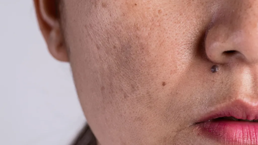
Melasma is a common facial skin pigmentation spot. It not only destroys the evenness and radiance of the skin, but may also cause low self-esteem and anxiety in patients, especially in social situations.Dermoscopy can clearly observe the characteristics of melasma such as the distribution of pigment granules, morphology and borders, which helps doctors to determine…
Melasma is a common facial skin pigmentation spot. It not only destroys the evenness and radiance of the skin, but may also cause low self-esteem and anxiety in patients, especially in social situations.
Dermoscopy can clearly observe the characteristics of melasma such as the distribution of pigment granules, morphology and borders, which helps doctors to determine the type and severity of melasma more accurately. Compared with the traditional naked eye observation, dermoscopy can reduce the errors caused by subjective judgement and improve the accuracy of diagnosis.
Overview of Melasma
Melasma is a common hyperpigmented skin disorder. It has a slow course and often has no other discomfort, but it seriously affects the skin’s aesthetics. The causes of melasma are complex and diverse, involving heredity, ultraviolet radiation, sex hormones, air pollution, mental and psychological factors, poor quality cosmetics, heat exposure and systemic diseases.
Melasma can be classified into the following types: epidermal, dermal, mixed and inflammatory. Ultraviolet rays are an important factor affecting melasma with prolonged exposure to sunlight, especially without cover. UV rays can damage the skin’s barrier function and cause an increase in melanocytes, which in turn leads to melasma.
Principles of Dermoscopy
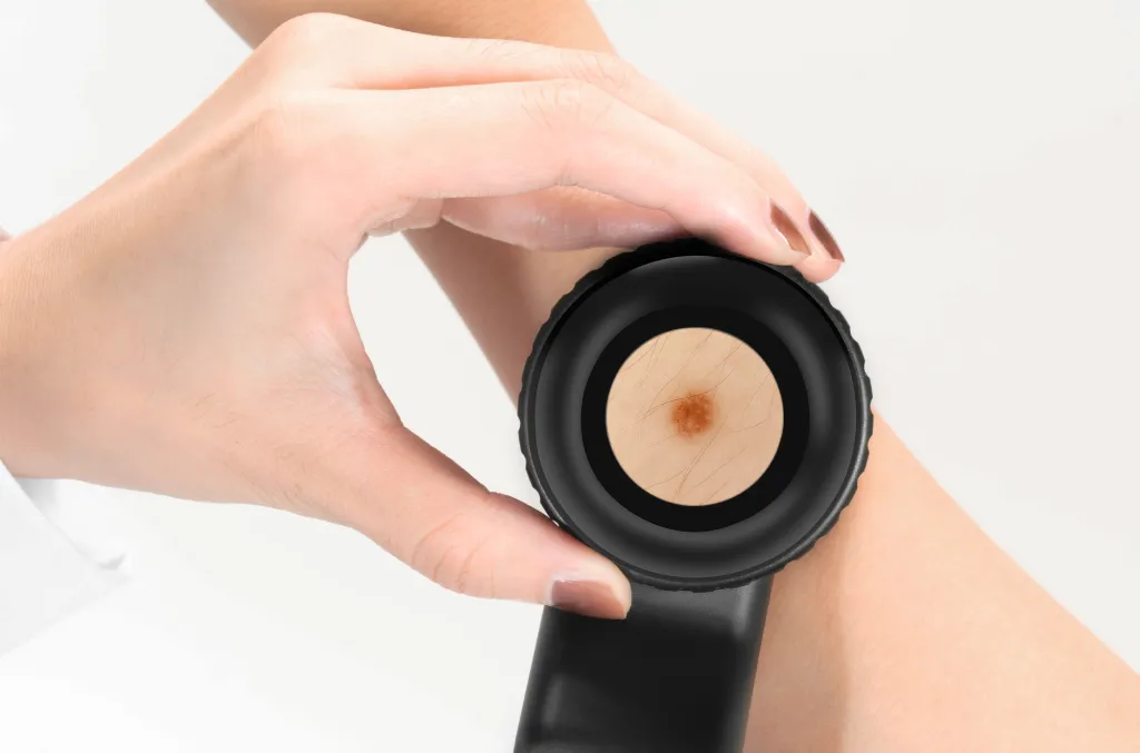
Dermoscopy is a non-invasive procedure that does not require cutting or sampling of the skin and therefore does not cause pain or trauma to the patient. Dermoscopy is able to magnify and visualise skin structures that are difficult to observe with the naked eye, enabling doctors to more accurately observe and analyse the characteristics of skin lesions.
Dermoscopy is able to clearly display the morphology, colour and structural features of skin pigmented lesions, which helps doctors to more accurately determine the nature and type of lesions. For example, when distinguishing between melanoma and benign nevus, a dark pigmented nevus will show obvious uneven pigmentation and different shades under dermoscopy.
Dermoscopy Procedure for Melasma
Before the start of dermoscopy, the patient needs to clean the skin surface to be examined in advance, and inform the doctor if there is any local inflammation or breakage, so that the doctor can assess the suitability of dermoscopy. During the examination, the doctor will put the probe of the dermatoscope on the surface of the skin lesion that needs to be observed, keeping a good distance between the probe and the skin. Adjust the light source and magnification of the device until the picture is clear. The doctor will select representative areas of melasma for examination, including the edges and centre of the lesion as well as the surrounding normal skin areas for comparative analysis.
Dermoscopic Features of Melasma
Typical dermoscopic manifestations of melasma are pseudopigmented networks of light to dark brown or greyish-blue patches, dots, globules, and curved or ring-shaped structures. Other dermoscopic features include capillary dilatation and thickening and darkening of cuii hair.
Dermoscopy can be used to differentiate melasma from other facial pigmented disorders. The following are the differences between melasma and other pigmented lesions:
Freckles: appear dermoscopically as round or oval light brown patches and brown punctate structures with well-defined borders.
Riehl’s melanosis: dermoscopically it shows a pseudopigmented network and blue-grey globules, capillary dilatation is seen in almost all lesions, and scaling is visible in some lesions.

Tips for Analysing Dermoscopic Images of Melasma
Observing the distribution of pigment in dermoscopic images allows one to determine the level and extent of pigment deposition. For example, when melanin when located at the true epidermal junction, it appears dermoscopically as dark or light brown. Observing the clarity of the borders of areas of pigmentation can help determine the type and staging of melasma. For example, the epidermal type of melasma usually has clear borders, whereas the dermal or mixed type may have fuzzy borders.
Regular monitoring with dermoscopy during the course of treatment allows for observation of changes in melasma, including reduction in pigmentation, change in pattern, and definition of borders. These changes can be used as a basis for assessing the effectiveness of the treatment.
Diagnosis and Differential Diagnosis of Melasma
To accurately differentiate dermal melasma from epidermal melasma, more specialised investigations such as histopathology are usually required. Dermoscopy can serve as a pre-diagnostic aid by optically magnifying the lesions. Melasma does not usually turn into melanoma. Melasma is a pigmentation. Melanoma is a malignant tumour caused by an abnormal proliferation of melanocytes. Although both involve hyperpigmentation, their pathological mechanisms and prognosis are very different.

Dermoscopy in Melasma Treatment
With dermoscopy, doctors can see the minuscule structures of the skin, determining the type and severity of skin lesions. Epidermal melasma may require a superficial peel, whereas dermal melasma may require a deeper laser treatment or medication.
Dermoscopy images at regular intervals allow clinicians to monitor the activity of the lesions and their treatment response, and to anticipate treatment outcomes. During the course of treatment, subsequent melasma dermoscopic images would show a progressive improvement in the degree of hyperpigmentation, with even distribution of pigment and clearer borders of the areas of hyperpigmentation over time.
Modern Methods of Melasma Treatment combined with Dermoscopy
The shape, extent and depth of the skin lesion dermoscopically is made so clear which helps the doctor to figure out what area needs to be specifically targeted for laser treatment. This allows to make the laser treatment more accurate sparing the adjacent normal skin. With the application of dermoscopy, the microstructure of the skin lesion can be observed, which will lead the doctor to diagnose the severity of the lesion and, therefore to choose the correct topical medication.
During treatment, dermoscopy can be used to dynamically monitor changes in the lesion. If the treatment is ineffective or the condition deteriorates, the doctor can optimise the treatment plan in time.
Melasma Patient Education and Self-monitoring
Patient understanding of the causes and typical symptoms of melasma will help patients to detect changes in their condition and take appropriate treatment measures. If you wish to use dermatoscope for home skin inspection, then IBOOLO dermatoscope will be your first choice, we will teach you how to operate it with DE-4100 as an example. First the area of however the skin to be examined is cleaned up, after which the dermatoscope magnet is connected to the mobile phone and the camera of the mobile phone is activated. Focus the image until it’s clear, then take a picture to document the event.
Limitations and challenges of dermoscopy
The patient’s skin colour, skin texture and skin type can affect dermoscopic observation. Inadequate or uneven lighting may result in blurred or distorted dermoscopic images. Prior to dermoscopy, patients should carefully clean the skin lesion area to ensure that there is no makeup or skin care product residue. Choose a place where the light is bright and even to perform the dermoscopy.
The key role of dermoscopy in the diagnosis and management of melasma
Dermoscopy contributes to a better visualisation of the deeper components of the skin and assists in the accurate diagnosis of a melasma, including its type, size and distribution, in order to establish more targeted treatment. Doctors can differentiate melasma with other pigmented skin diseases using dermoscopic image analysis and can avoid misdiagnosis after that. Dermoscopy is a promising approach to improving the diagnostic accuracy and efficiency of melasma and other skin diseases with continuous education and technological innovation as important drivers for the continuous development of dermoscopy.
Melasma Dermoscopy: Guide to Diagnosis & Treatment - IBOOLO
Melasma dermoscopy helps diagnose and monitor pigmentation disorders by revealing hidden skin patterns. Learn how it improves accuracy and guides treatment for better outcomes. Ideal for dermatologists and patients.
Melasma Dermoscopy: A Comprehensive Guide to Diagnosis, Treatment, and Monitoring
Melasma dermoscopy is a transformative tool in dermatology, offering a non-invasive way to diagnose and monitor this challenging pigmentation disorder. By magnifying the skin's surface, dermoscopy reveals intricate patterns and structures invisible to the naked eye, helping clinicians differentiate melasma from other conditions like post-inflammatory hyperpigmentation or solar lentigines. This guide explores the clinical applications of dermoscopy, from identifying epidermal vs. dermal melasma to tracking treatment progress. Whether you're a dermatologist refining your diagnostic skills or a patient seeking clarity, learn how melasma dermoscopy enhances accuracy, guides personalized treatment, and improves outcomes.
What is Melasma Dermoscopy: A Definitive Guide for Clinicians and Patients
Melasma dermoscopy is a non-invasive diagnostic technique that allows clinicians to examine skin lesions associated with melasma in greater detail. Unlike traditional visual inspection, dermoscopy uses a specialized device called a dermatoscope to magnify the skin's surface and reveal patterns that are invisible to the naked eye. This method is particularly useful for differentiating melasma from other pigmentation disorders, such as post-inflammatory hyperpigmentation or solar lentigines.
For patients, understanding melasma dermoscopy can provide clarity about their condition. It helps them grasp the underlying causes of their pigmentation issues, such as the distribution of melanin in the epidermis or dermis. For clinicians, this technique is invaluable for accurate diagnosis and treatment planning, as it provides insights into the depth and severity of the pigmentation.
Why is the Structure of Modern Dermoscopes Essential for Melasma Evaluation?
Modern dermoscopes are designed with advanced features that make them indispensable for melasma evaluation. These devices typically include:
Polarized and Non-Polarized Light Modes: Polarized light reduces surface reflection, allowing clinicians to see deeper into the skin, while non-polarized light highlights superficial structures.
High-Resolution Magnification: Most dermoscopes offer 10x to 20x magnification, enabling detailed visualization of melanin patterns.
Cross-Polarization Filters: These filters enhance contrast, making it easier to identify subtle pigmentation changes.
Portable and Ergonomic Designs: Modern dermoscopes are lightweight and easy to use, making them suitable for both clinical and field settings.
The combination of these features ensures that clinicians can accurately assess melasma, distinguishing it from other skin conditions with similar presentations.
How Does Clinical Workflow for Melasma Assessment Using Dermoscopy Work?
A structured clinical workflow is crucial for effective melasma assessment using dermoscopy. Here’s a step-by-step guide:
1. Patient History and Initial Examination: Begin by gathering the patient’s medical history and conducting a visual inspection of the affected areas.
2. Dermoscope Setup: Choose the appropriate light mode (polarized or non-polarized) based on the area being examined.
3. Image Capture: Use the dermoscope to capture high-resolution images of the pigmented lesions.
4. Pattern Analysis: Analyze the dermoscopic images for specific patterns, such as reticular, globular, or homogeneous pigmentation.
5. Documentation: Record findings in the patient’s medical records for future reference and treatment planning.
6. Treatment Recommendations: Based on the dermoscopic findings, recommend targeted treatments such as topical agents, chemical peels, or laser therapy.
This systematic approach ensures a thorough and accurate evaluation of melasma.
How Does Dermoscopy Reveal Melasma Patterns Invisible to the Naked Eye?
Dermoscopy works by illuminating the skin with controlled light and magnifying the reflected image. This process reveals intricate patterns and structures that are otherwise invisible. For melasma, dermoscopy can identify:
Epidermal vs. Dermal Pigmentation: Epidermal melasma appears as brown patches with well-defined borders, while dermal melasma shows a bluish-gray hue.
Vascular Patterns: Dermoscopy can highlight underlying blood vessels, which may indicate inflammation or vascular involvement in melasma.
Pigment Network: A reticular pattern of pigmentation is often visible, helping clinicians differentiate melasma from other conditions.
By uncovering these details, dermoscopy provides a deeper understanding of melasma’s characteristics, guiding more effective treatment strategies.
What Are the Proper Dermoscopy Techniques for Accurate Melasma Evaluation?
Accurate melasma evaluation requires adherence to proper dermoscopy techniques. Here are some key practices:
Consistent Lighting: Ensure the examination area is well-lit, and use the dermoscope’s built-in light source for consistent illumination.
Steady Hand Positioning: Hold the dermoscope firmly to avoid blurring the image.
Appropriate Magnification: Start with lower magnification (10x) to get an overview, then increase to 20x for detailed analysis.
Use of Contact Fluid: Apply a thin layer of gel or alcohol to the skin to reduce air gaps and improve image clarity.
Systematic Scanning: Move the dermoscope methodically across the lesion to ensure all areas are examined.
By following these techniques, clinicians can achieve reliable and reproducible results in melasma evaluation.
Can Dermoscopy Improve Melasma Treatment Outcomes?
Yes, dermoscopy can significantly improve melasma treatment outcomes by providing detailed insights into the condition’s characteristics. For example:
Targeted Therapy: By identifying the depth of pigmentation (epidermal vs. dermal), clinicians can tailor treatments more effectively.
Progress Monitoring: Dermoscopy allows for precise tracking of treatment progress, enabling adjustments as needed.
Patient Education: Visualizing the condition through dermoscopy helps patients understand their treatment plan and adhere to it.
What Are the Applications of Dermoscopy in Different Types of Melasma?
Dermoscopy is a versatile tool that can be applied to various types of melasma, each with distinct characteristics. Here’s how it aids in their evaluation:
1. Epidermal Melasma:
Dermoscopy reveals well-defined, brown pigmentation with a reticular or network-like pattern.
It helps confirm the superficial nature of the pigmentation, guiding the use of topical treatments like hydroquinone or retinoids.
2. Dermal Melasma:
This type shows a bluish-gray hue under dermoscopy due to melanin deposition in the deeper layers of the skin.
The technique helps identify the need for more aggressive treatments, such as laser therapy or chemical peels.
3. Mixed Melasma:
Dermoscopy can distinguish between epidermal and dermal components, providing a comprehensive view of the condition.
This dual insight allows for a combination of treatments tailored to both superficial and deep pigmentation.
By addressing the specific type of melasma, dermoscopy enhances diagnostic accuracy and treatment efficacy.
How Does Dermoscopy Accuracy Compare in Melasma vs. Other Facial Pigmentation Disorders?
Dermoscopy is highly accurate in differentiating melasma from other facial pigmentation disorders, such as post-inflammatory hyperpigmentation (PIH) or solar lentigines. Here’s how it compares:
Melasma:
- Shows a reticular or network-like pigmentation pattern.
- Often accompanied by telangiectasia (visible blood vessels) in chronic cases.
Post-Inflammatory Hyperpigmentation (PIH):
- Displays irregular, patchy pigmentation without a distinct network.
- May show signs of residual inflammation or scarring.
Solar Lentigines:
- Appears as well-defined, dark brown spots with a homogeneous pattern.
- Lacks the vascular changes often seen in melasma.
Dermoscopy’s ability to highlight these subtle differences makes it a superior tool for accurate diagnosis and treatment planning.
What Key Features Should You Look for in Dermoscopes for Melasma Assessment?
When selecting a dermoscope for melasma assessment, consider the following features to ensure optimal performance:
1. Polarized and Non-Polarized Light Modes:
Polarized light reduces glare and reveals deeper structures, while non-polarized light highlights surface details.
2. High Magnification (10x to 20x):
Allows for detailed visualization of pigmentation patterns and vascular structures.
3. Cross-Polarization Filters:
Enhances contrast, making it easier to identify subtle pigmentation changes.
4. Portability and Ergonomics:
Lightweight, handheld designs are ideal for clinical use.
5. Image Capture Capabilities:
Built-in cameras or smartphone compatibility for documenting findings.
Investing in a dermoscope with these features ensures accurate and efficient melasma evaluation.
Why Are Quality Standards Important in Dermoscopic Evaluation of Melasma?
Quality standards in dermoscopic evaluation are critical for ensuring consistent and reliable results. These standards include:
Standardized Imaging Protocols:
- Using consistent lighting, magnification, and contact fluids to minimize variability.
Training and Certification:
- Clinicians should undergo proper training to interpret dermoscopic patterns accurately.
Calibration of Equipment:
- Regular maintenance and calibration of dermoscopes to ensure optimal performance.
Documentation and Reporting:
- Detailed recording of findings to track progress and guide treatment decisions.
Adhering to these standards enhances the credibility of dermoscopic evaluations and improves patient outcomes.
What Precautions Should Be Taken When Using Dermoscopy for Melasma in Different Skin Types?
Dermoscopy is a safe and effective tool, but certain precautions are necessary when evaluating melasma in different skin types:
1. Fitzpatrick Skin Types IV-VI (Darker Skin Tones):
Use polarized light to reduce surface reflection and improve visualization.
Avoid excessive pressure, which can alter pigmentation patterns.
2. Fitzpatrick Skin Types I-III (Lighter Skin Tones):
Non-polarized light may be more effective for highlighting superficial pigmentation.
Be cautious of overexposure to light, which can cause discomfort.
3. General Precautions:
Clean the dermoscope and skin surface thoroughly to avoid contamination.
Use a thin layer of contact fluid to improve image clarity without distorting the skin’s appearance.
What is a Maintenance Guide for Dermoscopes Used in Pigmentation Clinics?
Proper maintenance of dermoscopes is essential to ensure their longevity and accuracy in pigmentation clinics. Here’s a step-by-step guide to keeping your device in optimal condition:
1. Daily Cleaning:
Wipe the lens and body of the dermoscope with a soft, lint-free cloth after each use.
Use a mild disinfectant to clean the contact plate, avoiding harsh chemicals that could damage the lens.
2. Weekly Inspection:
Check for scratches or cracks on the lens that could affect image quality.
Ensure the light source is functioning correctly and replace bulbs if necessary.
3. Monthly Calibration:
Verify the magnification and lighting settings to ensure consistent performance.
Test the device on a standardized skin model to confirm accuracy.
4. Storage Tips:
Store the dermoscope in a protective case to prevent dust accumulation and physical damage.
Keep it in a dry, temperature-controlled environment to avoid moisture-related issues.
By following this maintenance routine, clinics can extend the lifespan of their dermoscopes and maintain high diagnostic standards.
How Can Digital Dermoscopy Track Melasma Treatment Progress Over Time?
Digital dermoscopy is a powerful tool for monitoring melasma treatment progress. Here’s how it works:
1. Baseline Imaging:
Capture high-resolution dermoscopic images of the affected areas before starting treatment.
2. Regular Follow-Ups:
Take images at scheduled intervals (e.g., every 4-6 weeks) to track changes in pigmentation patterns.
3. Software Analysis:
Use specialized software to compare images and quantify changes in pigmentation intensity, distribution, and depth.
4. Adjusting Treatment Plans:
Based on the progress observed, modify treatment strategies to address persistent or new pigmentation.
Digital dermoscopy provides objective, visual evidence of treatment efficacy, helping both clinicians and patients stay informed and motivated.
What Do Dermoscopic Patterns in Melasma Reveal About Treatment Options?
Dermoscopic patterns in melasma offer valuable insights into the condition’s nature and guide treatment decisions. Key patterns include:
1. Reticular Pattern:
Indicates epidermal melasma, which responds well to topical treatments like hydroquinone or retinoids.
2. Bluish-Gray Hue:
Suggests dermal melasma, often requiring more intensive therapies such as laser treatment or chemical peels.
3. Telangiectasia (Visible Blood Vessels):
Points to vascular involvement, which may benefit from treatments targeting blood vessels, such as pulsed dye laser.
4. Homogeneous Pigmentation:
Common in mixed melasma, requiring a combination of superficial and deep treatment approaches.
By analyzing these patterns, clinicians can tailor treatments to the specific characteristics of the patient’s melasma.
How to Differentiate Melasma from Other Pigmentary Disorders Using Dermoscopy?
Dermoscopy is a key tool for distinguishing melasma from other pigmentary disorders. Here’s how to identify each condition:
1. Melasma:
Shows a reticular or network-like pigmentation pattern.
Often accompanied by telangiectasia in chronic cases.
2. Post-Inflammatory Hyperpigmentation (PIH):
Displays irregular, patchy pigmentation without a distinct network.
May show signs of residual inflammation or scarring.
3. Solar Lentigines:
Appears as well-defined, dark brown spots with a homogeneous pattern.
Lacks the vascular changes often seen in melasma.
4. Lichen Planus Pigmentosus:
Presents as gray-brown pigmentation with a granular or speckled pattern.
By recognizing these distinct features, clinicians can accurately diagnose melasma and rule out other conditions.
How Does Dermoscopy Assess Melasma Depth and Treatment Prognosis?
Dermoscopy provides critical information about melasma depth, which directly impacts treatment prognosis. Here’s how it works:
1. Epidermal Melasma:
Appears as brown patches with a reticular pattern under dermoscopy.
Indicates a better prognosis, as superficial pigmentation responds well to topical treatments.
2. Dermal Melasma:
Shows a bluish-gray hue, suggesting deeper melanin deposition.
Often has a poorer prognosis due to resistance to topical therapies, requiring advanced treatments like lasers.
3. Mixed Melasma:
Displays both epidermal and dermal patterns, indicating a variable prognosis.
Treatment success depends on addressing both superficial and deep pigmentation.
How to Integrate Dermoscopy into Your Melasma Treatment Protocol
Integrating dermoscopy into your melasma treatment protocol enhances diagnostic accuracy and treatment outcomes. Here’s a step-by-step approach:
1. Initial Assessment:
Use dermoscopy during the first consultation to evaluate the extent and depth of pigmentation.
Document baseline dermoscopic images for future comparison.
2. Diagnosis Confirmation:
Analyze dermoscopic patterns to differentiate melasma from other pigmentary disorders.
Identify specific features like reticular patterns or telangiectasia to confirm melasma type (epidermal, dermal, or mixed).
3. Treatment Planning:
Tailor treatments based on dermoscopic findings. For example, epidermal melasma may respond to topical agents, while dermal melasma might require laser therapy.
4. Progress Monitoring:
Schedule follow-up dermoscopic evaluations to track treatment efficacy.
Adjust treatment plans based on changes in pigmentation patterns.
By incorporating dermoscopy at every stage, clinicians can provide more precise and effective care for melasma patients.
How to Communicate Dermoscopic Findings to Melasma Patients
Effective communication of dermoscopic findings helps patients understand their condition and treatment plan. Here’s how to explain dermoscopic results clearly:
1. Visual Aids:
Show patients their dermoscopic images and highlight key features, such as pigmentation patterns or vascular changes.
2. Simplified Language:
Avoid medical jargon. For example, explain “reticular pattern” as a “net-like pigmentation” or “telangiectasia” as “visible blood vessels.”
3. Relate to Treatment:
Connect findings to the treatment plan. For instance, “The bluish-gray color means the pigmentation is deeper, so we’ll need stronger treatments like lasers.”
4. Set Realistic Expectations:
Use dermoscopic images to explain the expected timeline and potential outcomes of treatment.
By making dermoscopic findings relatable, clinicians can build trust and encourage patient compliance.
Why Combine Wood's Lamp and Dermoscopy for Comprehensive Melasma Evaluation?
Combining Wood’s lamp and dermoscopy provides a more comprehensive evaluation of melasma. Here’s how each tool contributes:
1. Wood’s Lamp:
Highlights epidermal pigmentation with a fluorescent glow, making it easier to identify superficial melasma.
Helps distinguish melasma from other conditions like fungal infections or vitiligo.
2. Dermoscopy:
Reveals detailed pigmentation patterns and vascular changes, aiding in the assessment of both epidermal and dermal melasma.
Provides high-resolution images for documentation and progress tracking.
3. Synergistic Benefits:
Wood’s lamp offers a quick overview, while dermoscopy provides in-depth analysis.
Together, they ensure a thorough evaluation, improving diagnostic accuracy and treatment planning.
This combination is particularly useful for complex or treatment-resistant melasma cases.
How Does Teledermoscopy Work for Remote Melasma Assessment and Monitoring?
Teledermoscopy enables remote evaluation and monitoring of melasma, making it a valuable tool for patients who cannot visit clinics in person. Here’s how it works:
1. Image Capture:
Patients or local healthcare providers use a portable dermoscope to capture high-quality images of the affected areas.
2. Data Transmission:
Images are securely uploaded to a telemedicine platform for review by a dermatologist.
3. Remote Analysis:
Dermatologists analyze the images to assess pigmentation patterns, depth, and treatment progress.
4. Treatment Recommendations:
Based on the findings, clinicians provide tailored treatment advice or adjustments via virtual consultations.
Teledermoscopy ensures continuity of care, especially for patients in remote or underserved areas.
How to Become Proficient in Melasma Dermoscopy Interpretation: A Training Guide
Proficiency in melasma dermoscopy interpretation requires structured training and practice. Here’s a step-by-step guide:
1. Learn the Basics:
Understand the principles of dermoscopy and its applications in pigmentation disorders.
Familiarize yourself with common dermoscopic patterns in melasma, such as reticular, globular, and homogeneous pigmentation.
2. Hands-On Practice:
Use a dermoscope to examine melasma cases under supervision.
Compare your findings with established dermoscopic criteria to build confidence.
3. Case Studies and Workshops:
Participate in workshops or online courses focused on melasma dermoscopy.
Review case studies to learn how to differentiate melasma from other pigmentary disorders.
4. Continuous Learning:
Stay updated with the latest research and advancements in dermoscopy.
Join professional networks or forums to exchange knowledge and experiences.
Melasma dermoscopy is an essential technique for diagnosing and managing melasma, a common yet complex pigmentation disorder. This guide highlights how dermoscopy reveals detailed patterns, such as reticular pigmentation or bluish-gray hues, to differentiate melasma types (epidermal, dermal, or mixed) and tailor treatments accordingly. It also covers the importance of modern dermoscope features, structured clinical workflows, and proper techniques for accurate evaluation. Additionally, the guide explores the role of digital dermoscopy in tracking treatment progress and the integration of tools like Wood’s lamp for comprehensive assessment. By leveraging dermoscopy, clinicians can improve diagnostic precision, enhance patient communication, and achieve better treatment outcomes for melasma.


