


Article
Dermoscopy of Melanocytic Nevus
People usually call mole as melaocytic nevus in the medical field. In most cases, melanocytic nevus presents benign and harmless to the skin. While there is a very low rate to turn into a melanoma from melanocytic nevi. But melanocytic is very common, and it can happen in everywhere on the skin. Thus, it is…
People usually call mole as melaocytic nevus in the medical field. In most cases, melanocytic nevus presents benign and harmless to the skin. While there is a very low rate to turn into a melanoma from melanocytic nevi. But melanocytic is very common, and it can happen in everywhere on the skin. Thus, it is really important to examine,monitor and follow up the melanocytic nevus by dermoscopy regularly.
What is melanocytic nevus?
Melanocytic nevus or mole, as a benign tumor, is a type of pigment cells (melanocytic) surging in large quantity on the skin. Melanocytic nevus appear from birth, so it often is called congenital malenocytic nevi, for short as CMN. Though melanocytic nevi usually do not bring harm to health. In same cases, they will develop into melanoma due to some factors.
What are clinical features of melanocytic nevus?
Though melanocytic nevi is harmless to health, but it is very crucial to recognize them from melanoma. There are some general clinical features which can tell melanocytic nevi as such as: Melanocytic nevi usually appear as flat or slightly higher than surrounding skin. The color of melanocytic nevi is commonly tan, skin-colored, brown and black. Different from melanoma, melanocytic nevi has symmetrial structure. And its border is well-fined and smooth.
What causes melanocytic nevus?
Memanocytic nevus commonly exhibits from the birth of baby, some melanocytic nevi come by several factors, such as: a family history, genetic factors, sun exposure and low immune system.
Application of dermoscopy in the diagnosis of melanocytic nevus
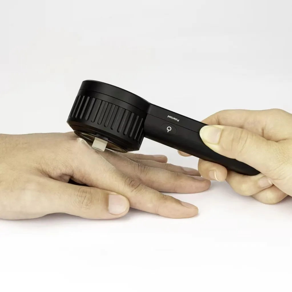
As an noninvasive technique, dermoscopy plays a very important role in skin lesions and skin diseases, including melanocytic nevus. Combining a powerful optical system and physical magnification, dermoscopy allows a deeper and enhanced visualization of the details of skin, such as its patterns, structures, colors and vessels. According to the distribution and numbers of pigmentation, dermoscopy helps dermatologists to get a clear and precise detection and diagnosis.
Dermoscopic features of benign melanocytic nevus
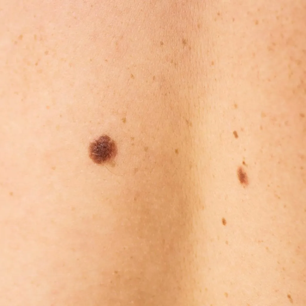
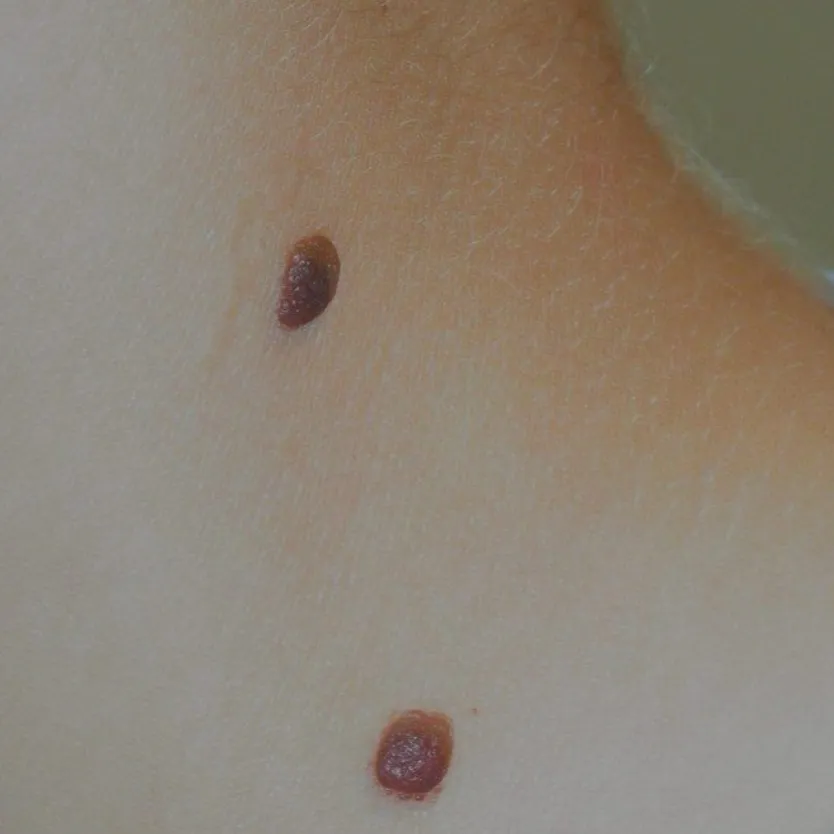
Dermoscopy can help dermatologists to identify and diagnose benign melanocytic nevus more accurately. There are several typical features of benign melanocytic nevus under a dermoscopy. Unlike melanoma, benign melanocytic nevus commonly with a asymmetrical shape and in large size. The structure of melanocytic nevus often is reticular, presenting brown or black globules aggregated by pigment network. Usually there are follicular openings or terminal hairs on the surface of the skin. Exhibiting parallel pattern on soles and palms of the skin.
Global structure and type of melanin nevus
The structure have different forms. Including well-defined, pigmented, round or oval lesions. As well,the size is tend to very small,like a coin or smaller. However, it still possible to appear big one. They maybe flat, flat peripherally and raised centrally, or entirely elevated. Fortunately, the elevated melanin nevus express a good “wobble sign” which can be shifted under the dermatoscope. Regarding the colors, that is common to see two or more which scatters average. Except for that, the obviously shape is Symmetry and uniformity. They can find out from anywhere on your body.
Identification of specific subtypes of melanocytic nevi
Ephelides:They are usually 1-3 mm in diameter but can be larger. They are usually light brown, darken in the summer, and fade without sun exposure.
Lentigo simplex:It’s the most common type of lentigo and can appear anywhere on the body, including areas that aren’t exposed to sunlight. Lentigo simplex is not caused by sun exposure and is not associated with systemic disease.
Solar lentigo: A solar lentigo is a flat, well-circumscribed patch. It can be round, oval or irregular in shape. Colour varies from skin-coloured, tan to dark brown or black, and size varies from a few millimetres to several centimetres in diameter.
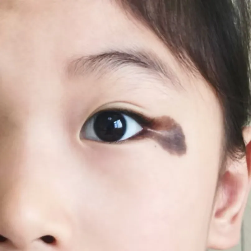
Congenital nevi: Congenital nevus melanogaster exists at the time of birth of infants. Congenital nevus melanogaster comes in various forms with different sizes, shapes and colors, and it can appear in any part of the body.
Congenital nevi consist of the following main patterns in dermoscopy: Honeycomb-like reticular pattern, lesions consisting of spherical patterns of varying sizes and homogenous patterns.
The difference of melanin nevus under clinical and dermatoscopy
Nevi of the palms and feet are mostly junctional nevi. The nevus cells of this kind of nevus are located at the junction of epidermis and dermis, usually small, with a diameter of 1-6 mm, smooth, hairless, flat or slightly elevated above the skin surface, with a color ranging from light brown to dark brown.
The pseudo-network feature of facial nevus melanogaster refers to the fact that facial nevi often appear dermoscopically as a circular grid of uniform size formed around the mouth of the hair follicle. Clinically, facial nevi may appear as flat or slightly elevated spots or plaques. Under dermoscopy, these nevi show a distinctive pseudopigmented reticular pattern.
Characteristics and evolution of halo naevi
Halo nevus may be the result of an autoimmune reaction, i.e., the autoimmune system kills melanocytes while accidentally attacking the surrounding normal skin pigment cells. Perineural white spots of halo nevus often appear as structureless hypopigmented spots under dermoscopy.
Pseudohalo naevus is a skin lesion that resembles a halo nevus but has a different mechanism of occurrence and pathologic features. Recognition of Pseudohalo naevus is mainly based on clinical presentation, dermoscopic examination and histopathologic examination.
The classification of melanocytic nevi and their histological relevance
Under dermoscopic examination, melanocytic nevi can be further subdivided into various patterns such as reticular, globular, homogeneous, and starburst types. The correlation between dermoscopic features and histologic changes corresponds briefly as follows.The reticular pattern represents a uniform distribution of nevus cells within the dermis, the spherical pattern indicates clumped aggregation of nevus cells, and the homogeneous pattern is associated with a uniform distribution of nevus cells without significant aggregation.
Clinical patterns and features of nevi and melanomas in children
Children’s melanocytic nevi commonly manifest clinically as black or dark brown spots, patches, or papules on the skin, varying in size, shape, and color. Under dermoscopy, these nevi can exhibit a reticular pattern, a homogeneous pattern, and vascular structures. Although relatively rare, the clinical morphology of pediatric melanoma is similar to that of adults. Dermoscopic features of pediatric melanoma may include asymmetry, irregular borders, and abnormal vascular patterns. Due to the high malignancy of pediatric melanoma, early detection, diagnosis, and treatment are crucial.
Monitoring and management of melanocytic nevi
Regular monitoring of melanocytic nevi can promptly detect changes in their morphology, color, or size, which may serve as early warning signals of malignancy. Dermoscopy, which magnifies and examines the fine structures and color variations on the skin’s surface, exhibits a high degree of accuracy in differentiating the benignity from malignancy of melanocytic nevi. It aids physicians in identifying abnormal pigment distribution and patterns, thereby enhancing their ability to distinguish between various skin lesions.
The majority of melanocytic nevi are benign, and typically do not require any special treatment if they remain stable in morphology and show no significant changes. However, if noticeable alterations occur in the size, shape, or color of a melanocytic nevus, such as enlargement, elevation, or uneven pigmentation, medical intervention is necessary, and prompt medical attention should be sought.


