Article

Dermoscopy of Lichenoid Keratosis

Lichenoid Keratosis occurs in middle-aged and elderly people, especially in sun-exposed areas such as the head, face, neck and dorsum of the hands. It is characterized by a wide variety of clinical manifestations, which can be easily confused with other similar lesions such as seborrheic keratosis, lichen planus and chronic lichenoid keratosis, leading to misdiagnosis…

Lichenoid Keratosis occurs in middle-aged and elderly people, especially in sun-exposed areas such as the head, face, neck and dorsum of the hands. It is characterized by a wide variety of clinical manifestations, which can be easily confused with other similar lesions such as seborrheic keratosis, lichen planus and chronic lichenoid keratosis, leading to misdiagnosis and underdiagnosis. Dermoscopy can clearly show the pigment granules in lichenoid keratosis lesions, which are diffusely distributed and coarse, a feature that helps to differentiate lichenoid keratosis from other similar lesions.


What Is Lichenoid Keratosis?
Lichenoid keratosis, also known as benign lichenoid keratosis or isolated lichen planus keratosis, is a benign keratotic skin disease with moss-like histologic changes. Its etiology is unclear and may be related to a number of factors, including genetic factors, abnormalities of the immune system, viral infections, and chronic ultraviolet exposure. Due to the relative infrequency of lichenoid keratosis, specific prevalence data are difficult to obtain, but the incidence is generally considered to be low.
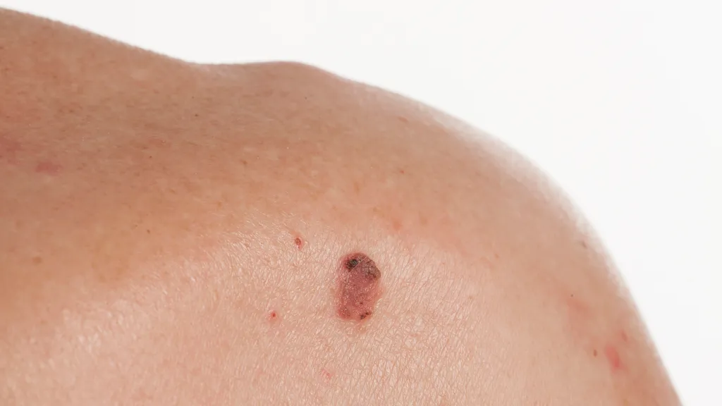
Lichenoid keratosis presents as single or multiple well-demarcated keratotic plaques, mostly red to dark red in color, some with uneven pigmentation. The surface of the lesions may be smooth or have wart-like protrusions, varying in size and usually ranging from 5 to 20 mm in diameter.

Lichenoid Keratosis
Lichenoid Keratosis


Clinical Distinction between Lichenoid Keratosis and Lichen Planopilaris
Lichenoid Keratosis: the color is mostly red to dark red, some have uneven pigmentation. The main manifestations are epidermal hyperkeratosis, thickening of the stratum spinosum and other moss-like changes.
Lichen Planopilaris: its lesions are mainly distributed around hair follicles, forming follicular papules or plaques, often accompanied by abnormalities and destruction of hair follicle structure.


Basics of Dermoscopy
Dermoscopes are mainly composed of a lens, a mirror body, and an adjustment knob. Before observing with a dermatoscope, clean the skin area to be examined. Then adjust the magnification and focusing distance of the dermatoscope as needed. Finally, place the dermatoscope on the skin area to be observed and observe the surface of the skin and the structure of the lower layers of the skin from different angles.
The high-resolution magnified images of dermoscopy can help doctors distinguish benign tumors from malignant skin cancer lesions. Moreover, dermoscopy is a non-invasive, visualized diagnostic tool for dermatological diseases, which does not traumatize the patient’s skin, reduces the patient’s nervousness and improves the patient’s cooperation.


Dermoscopic Features of Lichenoid Keratosis
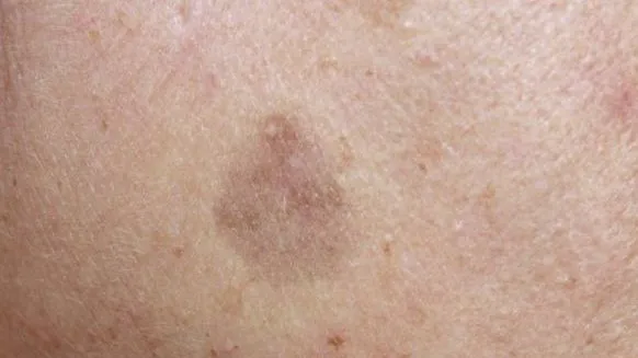
Lichenoid keratosis are often seen dermoscopically as pigment granules, which may be bluish-gray, brownish-red, or other colors. The pigment granules are diffusely distributed or may be focally distributed. They are usually less than 1 cm in diameter, flat or slightly elevated, and tend to have a smooth surface but occasionally scaly or warty. The lesions usually progress from an inflammatory phase (where some pink discoloration may occur) to a hyperpigmented phase (where the color of the lesion becomes grayish brown).

Dermoscopic Features of Lichenoid Keratosis


Lichenoid Keratosis and Other Lesions
Lichenoid keratosis, follicular lichenoid moss and malignant melanoma can be well differentiated by using dermoscopy, and the following are key points of their dermoscopic differentiation.
Lichenoid Keratosis usually has regular margins and lesions of various shapes. The pigmentation abnormalities in Lichenoid Keratosis may not be as dramatic as those of Malignant Melanoma, but some patients may still have deepening, hypopigmentation, or uneven pigmentation.
The distinguishing dermoscopic feature of follicular lichenoid moss is follicular involvement, such as hyperkeratosis of follicular openings, perifollicular erythema, or hyperpigmentation. The lesions may be covered with scales and are often associated with hyperkeratosis.
Malignant melanoma often appears dermoscopically as a mixture of colors with uneven distribution. The shape and color distribution of the lesions are often asymmetric and poorly demarcated from the surrounding normal skin.


Can Lichenoid Keratosis Become Cancerous?
Lichenoid Keratosis, a benign keratotic skin disease, does not usually transform into cancer. If the lesions are small and there are no obvious uncomfortable symptoms, they can be observed regularly and left untreated for the time being. If the lesion is large, or has obvious self-conscious symptoms, such as itching, pain, etc., affecting the patient’s daily life, then it can be considered for excision.

Lichenoid Keratosis
Lichenoid Keratosis


Dermoscopically Guided Clinical Decision Making
Utilizing dermoscopic findings for clinical decision-making, including biopsy and treatment selection, is a comprehensive process. When dermoscopic findings show skin lesions with malignant features or a high suspicion of malignancy, biopsy should be considered for definitive diagnosis. The choice of treatment, however, depends largely on the type of dermoscopic lesion. For benign lesions, observation and follow-up, topical medications, and physical therapy can be used for treatment. For malignant skin lesions, comprehensive treatment methods such as surgical excision, chemotherapy and radiotherapy should be chosen according to the severity of the disease.

Clinical Case Studies
Background
A middle-aged woman complained of a brown maculopapular rash on her shoulders for several years, which had recently increased in number and was accompanied by mild itching.
Clinical Examination:
The patient had multiple brown maculopapular rashes on the neck and shoulders, with rough and slightly flaky surfaces.
Dermoscopy:
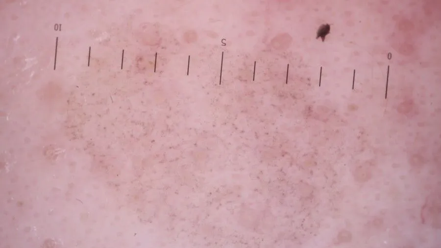
Dermoscopy revealed dense brownish-red pigmented granules on the surface of the lesions, which were diffusely or focally distributed. Worm-eaten changes were seen at the edges of some of the lesions, and hypopigmented areas were seen in the center of the lesions. The surface of the lesions is covered with a thin layer of scales and the skin texture is thickened.
Dermoscopic image interpretation:
Brownish-red pigmented granules help to differentiate between the different types of skin diseases. The lesions are covered with a thin layer of scales, suggesting hyperkeratosis and hypertrophy of the stratum spinosum. Wormy edges are a characteristic manifestation of Lichenoid Keratosis.

Dermoscopic Image
Dermoscopic Image


Treatment and Management of Lichenoid Keratosis
Topical treatments are one of the main therapeutic options for lichenoid keratosis, aiming to use medications that act directly on the lesions to relieve symptoms and promote skin recovery. Phototherapy uses specific wavelengths of ultraviolet light to irradiate the damaged area to promote hyperpigmentation and cell renewal, which can help improve mossy keratoses.
During the course of treatment, it is important to closely monitor the patient’s response to treatment, which includes observing changes in the lesions, assessing the degree of improvement in symptoms, and noting the presence of adverse reactions. And patients should be provided with appropriate scientific information and education, such as explaining the causes of lichenoid keratosis, instructing patients on how to use topical medications correctly, and encouraging patients to maintain a positive and optimistic mindset. All this is conducive to the recovery of patients.


Diagnostic Procedures for Dermoscopy
First, the patient needs to clean the skin surface to be examined, and the physician conducts a preliminary clinical examination to determine the possible types of diseases. The probe of the dermatoscope is gently applied to the skin surface to be observed, maintaining an appropriate distance. Then adjust the focus, carefully observe the skin lesions, the doctor will be based on their own experience to determine the type of skin disease. Dermoscopy is able to magnify and clearly display the fine structure of the skin surface, helping the physician to more accurately identify the characteristic changes of the skin lesion, thus improving the accuracy of diagnosis.

Share this article

0

No products in the cart.

Have questions on gear or your order?

Our Gear Guides are here to help! Get personal advice from pro creatives

Name
Subject
Email address
How can we help?

Instant Answers

How can dermoscopy images be captured?

Dermoscopy images can be captured and stored in different ways, such as: • Using a smartphone or tablet with dermoscopic adapter, which consisted in the package.• Using a digital camera

Dermoscopy images can be captured and stored in different ways, such as:

• Using a smartphone or tablet with dermoscopic adapter, which consisted in the package.
• Using a digital camera with dermoscopic adapter, there’s 49mm screw size camera adapter available to order now.

Compatible phone/tablet models:
All iPhone models, 95% Android phones, 90% tablet. For phone/tablet size in 5.25-14mm

Compatible camera models:
All camera with built 49mm filter screw, such as Canon EOS 70D, 80D, 90D; Canon EOS R7, R10, R50, R100; Canon M100, M200, M50, Mark II; Canon G7X Mark III, Sony ZV-1

How can I connect my phone to my dermatoscope?

There’s universal phone adapter for all our dermoscopes. Please check the installation procedure bellow or watch operation guide. Smartphone Connector (1) Place phone adapter screw in the center of smartphone’s

There’s universal phone adapter for all our dermoscopes. Please check the installation procedure bellow or watch operation guide.

Smartphone Connector

(1) Place phone adapter screw in the center of smartphone’s main camera.
(2) Screw magnet attachment on phone adapter.
(3) Put dermoscope’s back ring and magnet attachment together

Take The Best Images

You need to adjust the focus ring after the dermoscpe connected on smartphone to get the best images.

How can I clean my dermoscopy after usage?

Cleaning your dermoscopy after usage is important to prevent cross-contamination and infection. The cleaning method may vary depending on the type and model of your dermoscopy, so you should always

Cleaning your dermoscopy after usage is important to prevent cross-contamination and infection. The cleaning method may vary depending on the type and model of your dermoscopy, so you should always follow the manufacturer’s instructions. However, some general steps are:

• Turn off and disconnect your dermoscopy from any power source or device.

• Wipe off any visible dirt or debris from the dermoscopy with a soft cloth or tissue.

• Disinfect the dermoscopy with an alcohol-based wipe or spray, or a disinfectant solution recommended by the manufacturer. Make sure to cover all surfaces, especially the lens and contact plate.

• Let the dermoscopy air dry completely before storing it in a clean and dry place.

• Do not use abrasive or corrosive cleaners, solvents, or detergents that may damage the dermoscopy.

• Do not immerse the dermoscopy in water or any liquid, unless it is waterproof and designed for immersion.

You should clean your dermoscopy after each use, or at least once a day if you use it frequently. You should also check your dermoscopy regularly for any signs of damage or malfunction, and contact the manufacturer or service provider if needed.

Polarized VS Non-polarized Dermoscopy

A dermoscopy is a device that allows the examination of skin lesions with magnificationand illumination. By revealing subsurface structures and patterns that are not visible tothe naked eye. It can

A dermoscopy is a device that allows the examination of skin lesions with magnificationand illumination. By revealing subsurface structures and patterns that are not visible tothe naked eye. It can improve the diagnose accuracy of skin lesions, such as melanoma,basal cell carcinoma, seborrheic keratosis, etc.

There are two main types of dermoscopy: Non polarized and polarized dermoscopy.We’ve fitted most of our dermoscopys with polarized and non-polarized light. They canbe used in multiple skin structures.

Non-polarized contact Mode

In non-polarized mode, the instrument can provide information about the superficialskin structures, such as milia-like cysts, comedo-like openings, and pigment in theepidemis.

The dermoscopy requires applying a liquid such as mineral oil or alcohol to the skin andplacing the lens in contact with the skin. This reduces surface reflection and enhancesthe view of subsurface structures.

Image with non-polarized light (DE-3100)

Polarized contact Mode

In polarized mode, the instrument allows for visualization for deeper skin structures,such as blood vessels, collagen, and pigment in the dermis.

The dermoscopy does not need to be in contact with the skin or use any liquid. Theirpolarized light can help to eliminate surface reflection and allow visualization ofvascular structures.

Image with polarized light (DE-3100)

Polarized non-contact Mode

The dermoscopy can also use polarized light to examine the skin without direct contact.

In polarized non-contact mode, the instrument allows for examination infected areasand lesions that are painful for the patient, or the difficult to contact pigmented lesions,such as nails and narrow areas.

The contact plate should be removed in this mode, and it does not require applying aliquid to the skin. As it doesn’t require pressure or fluid application on the skin, it canalso avoid cross-contamination and infection risk.

Image in polarized non-contact mode (DE-3100)

How effectiveness is dermoscopy

Compared with visual inspection, the dermoscopy can be used to capture and store skin lesion photos, which play an important role in early skin cancer examination. The dermoscopy allows the

Compared with visual inspection, the dermoscopy can be used to capture and store skin lesion photos, which play an important role in early skin cancer examination.

The dermoscopy allows the examination of skin lesions with magnification and illumination. This can be greatly avoiding the factors that cause interference to visual detection. Such as lighting, skin color, hair and cosmetics.

Several studies have demonstrated that dermoscopy is useful in the identification of melanoma, when used by a trained professional.

It may improve the accuracy of clinical diagnosis by up to 35%
It may reduce the number of harmless lesions that are removed
In primary care, it may increase the referral of more worrisome lesions and reduce the referral of more trivial ones

A 2018 Cochrane meta-analysis published the accuracy of dermoscopy in the detection.

Table 1. Accuracy of dermoscopy in the detection of melanoma in adults
Detection Method Sensitivity, % Specificity, % Positive Likelihood Ratio NegativeLikelihood Ratio
Visual inspection alone (in person) 76 75 3.04 0.32
Dermoscopy with visual inspection (in person) 92 95 18 0.08
Image-based visual inspection alone (not in person) 47 42 0.81 1.3
Dermoscopy with image-based visual inspection (not in person) 81 82 4.5 0.23
ROC—receiver operating characteristic. *Estimated sensitivity calculated on the summary ROC curve at a fixed specificity of 80%.

As we can see, the dermoscope can improve the accuracy of diagnosis of skin lesions, especially melanoma.

Table 1. Accuracy of dermoscopy in the detection of melanoma in adults
Detection Method Sensitivity, % Specificity, % Positive Likelihood Ratio NegativeLikelihood Ratio
Visual inspection alone (in person) 79 77 3.4 0.27
Dermoscopy with visual inspection (in person) 93 99 93 0.07
Image-based visual inspection alone (not in person) 85 87 6.5 0.17
Dermoscopy with image-based visual inspection (not in person) 93 96 23 0.07
ROC—receiver operating characteristic. *Estimated sensitivity calculated on the summary ROC curve at a fixed specificity of 80%.

Characteristics of the dermatoscopic structure of the skin lesions include:

• Symmetry or asymmetry
• Homogeny/uniformity (sameness) or heterogeny (structural differences across the lesion)
• Distribution of pigment: brown lines, dots, clods and structureless areas
• Skin surface keratin: small white cysts, crypts, fissures
• Vascular morphology and pattern: regular or irregular
• Border of the lesion: fading, sharply cut off or radial streaks
• Presence of ulceration

There are specific dermoscopic patterns that aid in the diagnosis of the following pigmented skin lesions:

• Melanoma
• Moles (benign melanocytic naevus)
• Freckles (lentigos)
• Atypical naevi
• Blue naevi
• Seborrhoeic keratosis
• Pigmented basal cell carcinoma
• Haemangioma

Hot Search Terms