Article

Dermoscopy of Lichen Planopilaris

The clinical significance of lichen planopilaris is mainly reflected in the impact on patients’ quality of life. It can lead to patchy or diffuse baldness of the scalp, which not only affects the patient’s appearance, but may also have a negative impact on the patient’s mental health. Early and accurate diagnosis of lichen planopilaris can…

The clinical significance of lichen planopilaris is mainly reflected in the impact on patients’ quality of life. It can lead to patchy or diffuse baldness of the scalp, which not only affects the patient’s appearance, but may also have a negative impact on the patient’s mental health. Early and accurate diagnosis of lichen planopilaris can help to take timely treatment measures to slow down the progression of the disease and reduce the area of hair loss. Dermoscopy can clearly observe the characteristic lesions such as erythema, desquamation, follicular hyperkeratosis, and clustered spiny follicular papules around the hair follicles of patients with lichen planopilaris, which can provide an intuitive and accurate basis for the diagnosis of lichen planopilaris.


What Is Lichen Planopilaris?
Lichen Planopilaris, also known as trichophytic lichen planus, is a form of primary lymphocytic scarring alopecia that mainly effects the scalp. The exact cause of tinea versicolor is unknown, but it may be the result of a variety of issues including immune system abnormalities, genetic factors, fungus on your skin and emotional stress.
Lichen planopilaris mainly shows patchy alopecia or diffuse hair loss on the scalp, with active lesions at the margins of bald patches including perifollicular erythema and desquamation with follicular hyperkeratosis. Associated symptoms include pruritus and pain, which are various in severity.

Lichen Planopilaris
Lichen Planopilaris


Epidemiology and Classification of Lichen Planopilaris
The incidence of lichen planopilaris is relatively low worldwide, with a reported prevalence of about 1%. More often than men followed by women, this disease is present in the age bracket of 30 to 70 years old. Lichen planopilaris can be classified into the following types or subtypes based on their clinical presentation and pathologic features: classic lichen planopilaris, frontal fibrosing alopecia, and Graham-Little-Piccardi-Lasseur syndrome.


Dermoscopy in the Diagnosis of Lichen Planopilaris
Dermoscopy is an optically based dermatologic diagnostic tool that focuses on magnifying the surface of the skin through the use of an optical magnification system so that the physician can see the texture and details of the skin surface more clearly. Dermoscopy also improves the contrast and clarity of the image through the use of optical filters and color filters.
Lichen planopilaris is a chronic inflammatory skin disease that primarily affects the hair follicles of the scalp. It has some characteristic manifestations under dermoscopy, such as erythema around the hair follicles, punctate hemorrhage at the mouth of the hair follicles, and atrophy of the scalp, which can help doctors to make a differential diagnosis. Meanwhile, dermoscopy, as a non-invasive means of examination, can observe the characteristics of lesions without damaging the skin, reducing the pain and discomfort of patients.
Prior to dermoscopy, patients should avoid applying medications or cosmetics to the skin surface prior to the examination, and especially avoid applying sunscreen and other substances that may block light from the imaging. During the examination, the patient should try to cooperate with the examiner and assume the proper position so that the skin lesions can be fully exposed. After the examination, patients need to pay attention to keep the local skin clean and dry, and avoid scratching the affected area, so as not to cause skin infection.

Dermoscopy of Lichen Planopilaris


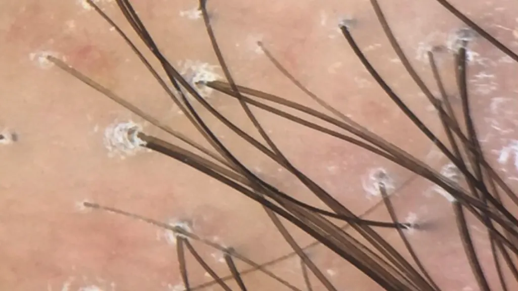
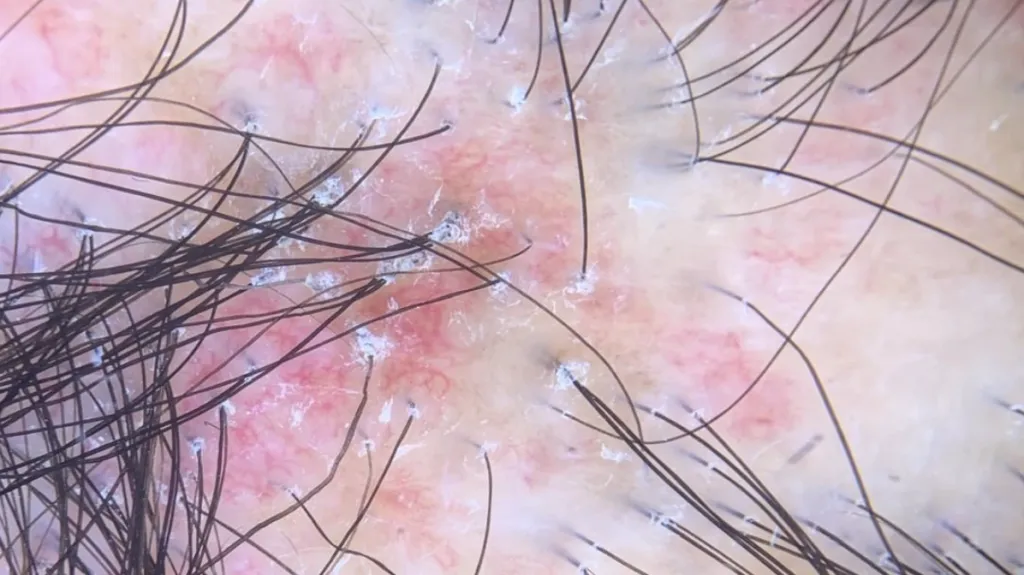
Dermoscopic Features of Lichen Planopilaris
Absence of follicular openings: In advanced lesions of Lichen Planopilaris, the follicular openings may be completely absent, resulting in visible patches of alopecia on the scalp.
Perifollicular erythema: In active lesions, perifollicular erythema can be observed, and these are usually accompanied by signs of desquamation and follicular hyperkeratosis.
Classic white and blue-gray spots: On dermoscopic examination, Lichen Planopilaris may exhibit irregular white spots between hair follicles and bluish-gray spots around hair follicles.
White scarred areas: Areas of white scarring usually form as a result of complete loss of hair follicles and fibrosis of the skin, and are typical of advanced stages of lichen planus.
Milky red areas: On dermoscopy, localized congestion or vasodilatation due to an inflammatory response may be observed, and these usually appear as red or pink areas.


Lichen Planopilaris and Other Alopecia Areata Disorders
Lichen Planopilaris differs significantly from other alopecia areata disorders (e.g., discoid lupus, frontal fibrotic alopecia) in terms of dermoscopic features. Dermoscopy is able to magnify the area of skin lesions, details of skin surface microstructure, hair shaft morphology, and capillaries, providing visual evidence for differential diagnosis.
Lichen Planopilaris: Dermoscopy reveals a marked inflammatory reaction around the hair follicle in the form of erythema, edema, or desquamation, and these changes are distributed around the hair follicle in a ring or target-shaped pattern. The follicular opening may become inconspicuous or disappear completely, and sometimes the follicular opening can be seen to be blocked by keratin plugs.
Discoid Lupus: The follicular opening is seen as a distinct red spot, which is often surrounded by a white halo. In advanced lesions, due to fibrosis of the dermis, white structureless areas are seen dermoscopically; these areas correspond to scar tissue in the dermis.
Frontal Fibrotic Alopecia: There is a marked reduction in the number of hair follicles in the frontal region, especially the reduction of coarse hairs. Erythema around the hair follicles may be present in the area of hair loss. The forehead skin may become smooth and tight, losing normal skin texture and elasticity.
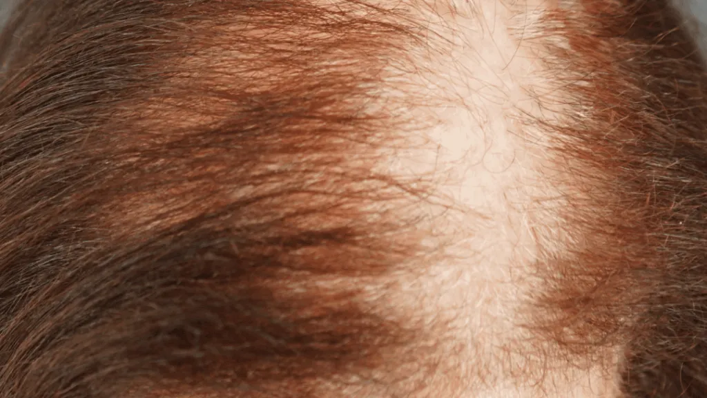

Case Study
Clinical and dermoscopic images of lichen planopilaris are shown below respectively.

Clinical Image of Lichen Planopilaris
Dermoscopic Images of Lichen Planopilaris


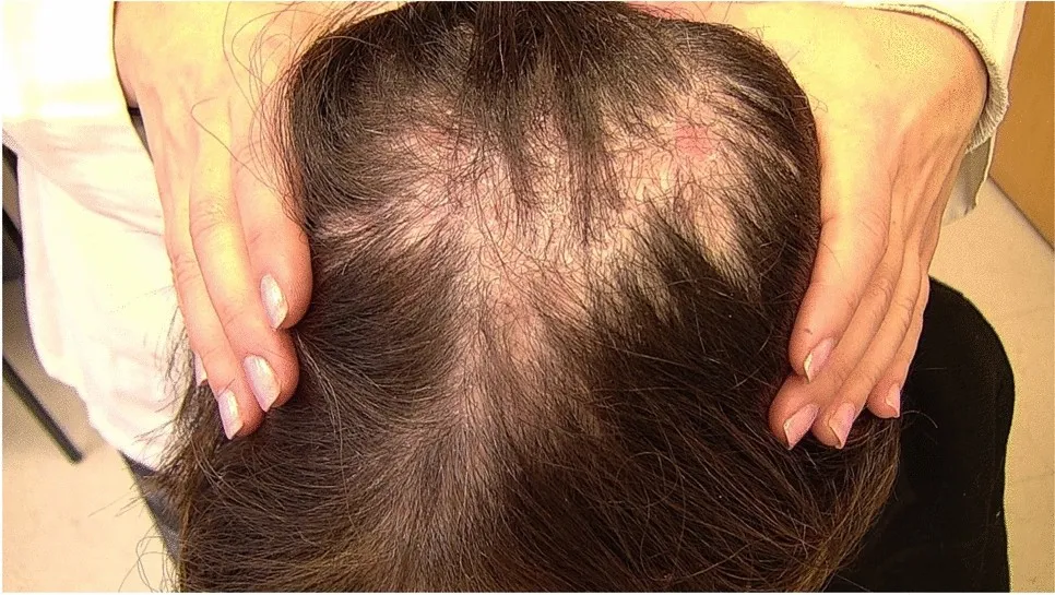
By comparing the clinical and dermoscopic images of lichen planopilaris we can see that the dermoscopic images show the fine structure of the follicular units more clearly. The dermoscopic image shows scales around the hair follicle as well as a hair tube pattern, and keratinous plugs are visible at the follicular openings, which are due to hyperkeratosis and blockage of the follicular openings with large amounts of keratinized material. Blue-gray spots around the hair follicles, forming target-like pigmentation, are typical features of lichen planopilaris, and the doctor can make a preliminary judgment of the disease based on them.
Dermoscopy is an important tool for initial diagnosis, but pathologic examination remains the gold standard for diagnosis. For patients with suspected LPP, pathologic examination should be performed as early as possible to confirm the diagnosis and reduce the possibility of misdiagnos.


Treatment and Management of Lichen Planopilaris
The treatment of lichen planopilaris mainly includes medication, physical therapy and surgery. Patients should eat more fresh vegetables, ensure enough sleep and face the disease with an optimistic attitude to help the disease recover. After treatment, doctors should closely observe whether the patient’s symptoms are reduced. According to the improvement of the patient’s rash, it will be categorized into four grades for evaluation: cured, obvious effect, effective and ineffective.
Lichen planopilaris is a chronic disease that requires long-term management and treatment. Patients should take the medication on time as prescribed by the doctor and should not stop taking the medication or change the dosage at will. Health education should be strengthened to improve patients’ knowledge of the disease and self-management ability.

Share this article

0

No products in the cart.

Have questions on gear or your order?

Our Gear Guides are here to help! Get personal advice from pro creatives

Name
Subject
Email address
How can we help?

Instant Answers

How can dermoscopy images be captured?

Dermoscopy images can be captured and stored in different ways, such as: • Using a smartphone or tablet with dermoscopic adapter, which consisted in the package.• Using a digital camera

Dermoscopy images can be captured and stored in different ways, such as:

• Using a smartphone or tablet with dermoscopic adapter, which consisted in the package.
• Using a digital camera with dermoscopic adapter, there’s 49mm screw size camera adapter available to order now.

Compatible phone/tablet models:
All iPhone models, 95% Android phones, 90% tablet. For phone/tablet size in 5.25-14mm

Compatible camera models:
All camera with built 49mm filter screw, such as Canon EOS 70D, 80D, 90D; Canon EOS R7, R10, R50, R100; Canon M100, M200, M50, Mark II; Canon G7X Mark III, Sony ZV-1

How can I connect my phone to my dermatoscope?

There’s universal phone adapter for all our dermoscopes. Please check the installation procedure bellow or watch operation guide. Smartphone Connector (1) Place phone adapter screw in the center of smartphone’s

There’s universal phone adapter for all our dermoscopes. Please check the installation procedure bellow or watch operation guide.

Smartphone Connector

(1) Place phone adapter screw in the center of smartphone’s main camera.
(2) Screw magnet attachment on phone adapter.
(3) Put dermoscope’s back ring and magnet attachment together

Take The Best Images

You need to adjust the focus ring after the dermoscpe connected on smartphone to get the best images.

How can I clean my dermoscopy after usage?

Cleaning your dermoscopy after usage is important to prevent cross-contamination and infection. The cleaning method may vary depending on the type and model of your dermoscopy, so you should always

Cleaning your dermoscopy after usage is important to prevent cross-contamination and infection. The cleaning method may vary depending on the type and model of your dermoscopy, so you should always follow the manufacturer’s instructions. However, some general steps are:

• Turn off and disconnect your dermoscopy from any power source or device.

• Wipe off any visible dirt or debris from the dermoscopy with a soft cloth or tissue.

• Disinfect the dermoscopy with an alcohol-based wipe or spray, or a disinfectant solution recommended by the manufacturer. Make sure to cover all surfaces, especially the lens and contact plate.

• Let the dermoscopy air dry completely before storing it in a clean and dry place.

• Do not use abrasive or corrosive cleaners, solvents, or detergents that may damage the dermoscopy.

• Do not immerse the dermoscopy in water or any liquid, unless it is waterproof and designed for immersion.

You should clean your dermoscopy after each use, or at least once a day if you use it frequently. You should also check your dermoscopy regularly for any signs of damage or malfunction, and contact the manufacturer or service provider if needed.

Polarized VS Non-polarized Dermoscopy

A dermoscopy is a device that allows the examination of skin lesions with magnificationand illumination. By revealing subsurface structures and patterns that are not visible tothe naked eye. It can

A dermoscopy is a device that allows the examination of skin lesions with magnificationand illumination. By revealing subsurface structures and patterns that are not visible tothe naked eye. It can improve the diagnose accuracy of skin lesions, such as melanoma,basal cell carcinoma, seborrheic keratosis, etc.

There are two main types of dermoscopy: Non polarized and polarized dermoscopy.We’ve fitted most of our dermoscopys with polarized and non-polarized light. They canbe used in multiple skin structures.

Non-polarized contact Mode

In non-polarized mode, the instrument can provide information about the superficialskin structures, such as milia-like cysts, comedo-like openings, and pigment in theepidemis.

The dermoscopy requires applying a liquid such as mineral oil or alcohol to the skin andplacing the lens in contact with the skin. This reduces surface reflection and enhancesthe view of subsurface structures.

Image with non-polarized light (DE-3100)

Polarized contact Mode

In polarized mode, the instrument allows for visualization for deeper skin structures,such as blood vessels, collagen, and pigment in the dermis.

The dermoscopy does not need to be in contact with the skin or use any liquid. Theirpolarized light can help to eliminate surface reflection and allow visualization ofvascular structures.

Image with polarized light (DE-3100)

Polarized non-contact Mode

The dermoscopy can also use polarized light to examine the skin without direct contact.

In polarized non-contact mode, the instrument allows for examination infected areasand lesions that are painful for the patient, or the difficult to contact pigmented lesions,such as nails and narrow areas.

The contact plate should be removed in this mode, and it does not require applying aliquid to the skin. As it doesn’t require pressure or fluid application on the skin, it canalso avoid cross-contamination and infection risk.

Image in polarized non-contact mode (DE-3100)

How effectiveness is dermoscopy

Compared with visual inspection, the dermoscopy can be used to capture and store skin lesion photos, which play an important role in early skin cancer examination. The dermoscopy allows the

Compared with visual inspection, the dermoscopy can be used to capture and store skin lesion photos, which play an important role in early skin cancer examination.

The dermoscopy allows the examination of skin lesions with magnification and illumination. This can be greatly avoiding the factors that cause interference to visual detection. Such as lighting, skin color, hair and cosmetics.

Several studies have demonstrated that dermoscopy is useful in the identification of melanoma, when used by a trained professional.

It may improve the accuracy of clinical diagnosis by up to 35%
It may reduce the number of harmless lesions that are removed
In primary care, it may increase the referral of more worrisome lesions and reduce the referral of more trivial ones

A 2018 Cochrane meta-analysis published the accuracy of dermoscopy in the detection.

Table 1. Accuracy of dermoscopy in the detection of melanoma in adults
Detection Method Sensitivity, % Specificity, % Positive Likelihood Ratio NegativeLikelihood Ratio
Visual inspection alone (in person) 76 75 3.04 0.32
Dermoscopy with visual inspection (in person) 92 95 18 0.08
Image-based visual inspection alone (not in person) 47 42 0.81 1.3
Dermoscopy with image-based visual inspection (not in person) 81 82 4.5 0.23
ROC—receiver operating characteristic. *Estimated sensitivity calculated on the summary ROC curve at a fixed specificity of 80%.

As we can see, the dermoscope can improve the accuracy of diagnosis of skin lesions, especially melanoma.

Table 1. Accuracy of dermoscopy in the detection of melanoma in adults
Detection Method Sensitivity, % Specificity, % Positive Likelihood Ratio NegativeLikelihood Ratio
Visual inspection alone (in person) 79 77 3.4 0.27
Dermoscopy with visual inspection (in person) 93 99 93 0.07
Image-based visual inspection alone (not in person) 85 87 6.5 0.17
Dermoscopy with image-based visual inspection (not in person) 93 96 23 0.07
ROC—receiver operating characteristic. *Estimated sensitivity calculated on the summary ROC curve at a fixed specificity of 80%.

Characteristics of the dermatoscopic structure of the skin lesions include:

• Symmetry or asymmetry
• Homogeny/uniformity (sameness) or heterogeny (structural differences across the lesion)
• Distribution of pigment: brown lines, dots, clods and structureless areas
• Skin surface keratin: small white cysts, crypts, fissures
• Vascular morphology and pattern: regular or irregular
• Border of the lesion: fading, sharply cut off or radial streaks
• Presence of ulceration

There are specific dermoscopic patterns that aid in the diagnosis of the following pigmented skin lesions:

• Melanoma
• Moles (benign melanocytic naevus)
• Freckles (lentigos)
• Atypical naevi
• Blue naevi
• Seborrhoeic keratosis
• Pigmented basal cell carcinoma
• Haemangioma

Hot Search Terms