


Article
Dermoscopy Magnification
The dermatoscope uses a specific wavelength light source and optical magnification to allow the physician to visualize subtle structures and pigmentation changes that are difficult to distinguish with the naked eye. This high-definition imaging capability gives dermoscopy a significant advantage in identifying benign and malignant tumors of the skin and diagnosing pigmentary, inflammatory and vascular…
The dermatoscope uses a specific wavelength light source and optical magnification to allow the physician to visualize subtle structures and pigmentation changes that are difficult to distinguish with the naked eye. This high-definition imaging capability gives dermoscopy a significant advantage in identifying benign and malignant tumors of the skin and diagnosing pigmentary, inflammatory and vascular diseases. Higher magnification facilitates better delineation of the subtle morphologic and color alterations in a skin lesion. These minute differences are often the difference in key to diagnosing skin diseases at all.
Fundamentals of Dermoscopy
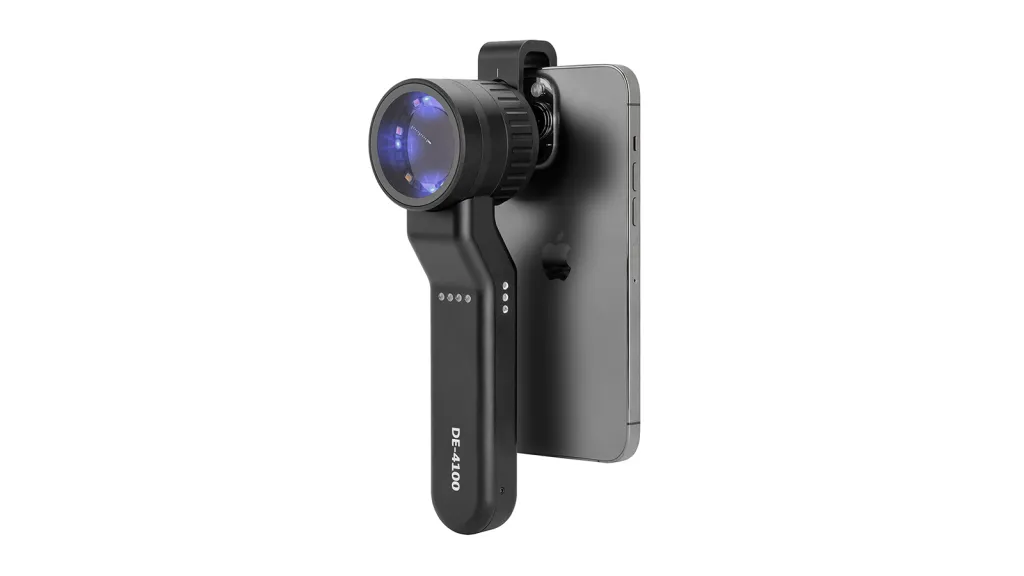
Dermatoscope operates primarily on the principles of optics, especially light scattering and transmission. Certain types of liquid media or polarized light could decrease the amount of scattered light by stratum corneum, and enable deeper penetration to skin surface layers not just within epithelium but in superficial dermis as well.
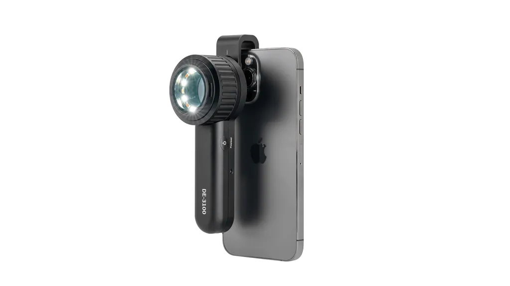
A dermatoscope is commonly composed of a handle, a lens and usually also contains polarizing filter. Dermoscopy, as the stethoscope of a dermatologist, has an irreplaceable role in clinical diagnosis for routine skin diseases. In that vein, dermoscopy has the Advantages of less easy to operate and detect with a short detection period well-suited for rapid screening/diagnose in outpatient clinics or other places.
The Importance of Magnification
If the magnification level is too low, crucial features like skin lesion contours and morphology or colour might not be observed properly by doctors leading to decreased performance in disease diagnosis. On the other hand, an excessively high magnification can lead to prolonged examination time. Additionally, due to its narrow field of view, shallow depth of field, and high demand for lighting conditions, such a high magnification is more suitable for specialized research and specific diagnostic needs.
Low magnification provides a large field of view, which is suitable for observing the overall morphology of the skin lesion, its distribution range, and its boundaries with the surrounding tissues. High magnification provides a very small field of view and is suitable for observing the microstructure of skin lesions and deep tissue changes.
Magnification Techniques for Dermoscopy
Dermoscopic magnification technology is mainly divided into non-contact magnification and contact magnification, both have their limitations and advantages.
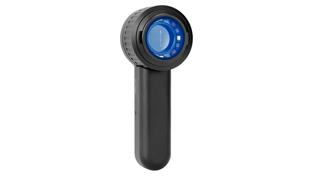
Non-contact dermoscopy does not directly contact the skin, avoiding the risk of cross-infection due to contact. Moreover, it eliminates the influence of external force on the pressure of blood vessels, which makes the observation result more accurate. However, due to the non-contact skin, the operator has a high level of skill and experience to ensure the accuracy of observation, and the cost of the product is relatively high.
Contact dermatoscopy is in direct contact with the skin, and can more clearly observe the subtle structure and changes on the skin surface, such as the boundary, morphology and color of the lesions. However, there is a risk of cross-infection between patients if the equipment is not thoroughly sterilized or operated properly.
Common skin lesions in dermoscopy
Common skin lesions under dermoscopy include pigmented lesions, non-pigmented skin lesions, hormone-dependent skin lesions, hair diseases, benign and malignant tumors. The need for magnification for different lesion types needs to be considered according to the type of lesion, severity of the disease and the purpose of observation. In practice, doctors will choose the appropriate magnification for observation based on specific circumstances and experience.
Magnification in the Diagnosis Skin Lesions
High-definition magnification of the dermatoscope clearly shows the fine structure of melanoma and basal cell carcinoma. Melanoma is characterized by its pigment network structure, blue-white curtain structure, stripe shape, and uniform dots. Basal cell carcinoma has blue-gray ovoid nests, blue-gray globules, spoke-like areas, and dendritic blood vessels.
Under low magnification dermoscopy, the physician can generally observe the overall shape and borders of the lesion. With increasing magnification, the doctor is able to observe more subtle features such as pigment network structure, blood vessel morphology, and cellular arrangement.
Clinical Case Study
Case Background
Ms. Li, a patient, presented with a persistently enlarged brown plaque on her face. On initial observation, the plaque had unclear borders and varied color shades, and was suspected to be a skin tumor.
Dermoscopic examination
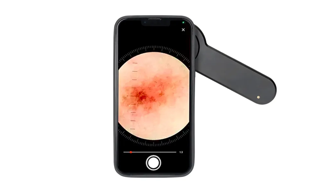
Under dermoscopy, the brown plaque on Ms. Li’s face showed obvious structural asymmetry, the presence of irregular punctate and reticular pigment distribution, and distorted vascular patterns.
The role of magnification
High magnification allows the doctor to observe subtle structural changes that are difficult to detect with the naked eye, such as the distribution pattern of pigment particles and the morphology of blood vessels. These subtle changes are critical in identifying the type of skin tumor and whether it is benign or malignant.
Correlation of dermoscopic images with final diagnosis
The image features observed by dermoscopy are highly consistent with the subsequent pathological diagnosis, and the final pathological diagnosis is skin tumor.
Dermoscopy Techniques
Ensure that the light source is moderately bright and stable during the dermoscopic examination. When holding the dermatoscope, the doctor should keep the arm stable and avoid shaking the hand, point the dermatoscope’s probe at the area under observation, and adjust the focus until the optimal field of view.
Hand-held dermatoscopes are generally capable of focusing and magnifying, allowing the physician to select the appropriate magnification according to the size of the lesions in the field of view. When the lesion in the field of view is too small, the magnification should be continued until the features of the lesion appear clearly.
Dermoscopy Procedure
Before the dermoscopy, the skin surface to be examined is cleaned, and then the probe of the dermoscopy is attached to the skin surface to be observed. Discuss the adjustment and application of magnification in the diagnostic process. A lower magnification may be used at first for initial observation of the lesions, followed by a gradual increase in magnification to visualize the skin’s microstructure in greater detail.
Education and Training
Training dermatoscope operators in the use of magnification is directly related to the accuracy and validity of the diagnosis of skin lesions. During the daily training, dermatoscopes can be used to demonstrate how to adjust the magnification according to the characteristics of the skin lesions by using hands-on demonstrations on actual cases. Provide samples or pictures of simulated skin lesions for the operator to practice repeatedly to familiarize himself with the observation effect under different magnifications. If you want to improve the diagnostic skills of dermoscopy, doctors need to continue to learn dermatology-related knowledge, participate in more clinical practice, and accumulate rich experience through actual operation.
Dermoscopy Technology in the Future
As a device that can magnify the structure of skin lesions, dermoscopy’s unique microscopic magnification and fluoroscopic capabilities greatly compensate for the limitations of naked eye observation. With the development of technology, digital dermoscopy is becoming more and more popular. Digital dermatoscopy not only inherits all the advantages of traditional dermatoscopy, but also transmits the images of skin lesions directly to the computer through digitalization technology, realizing instant processing and saving of images. In the future, with the use of artificial intelligence in the field of dermatoscopy, AI algorithms will learn and recognize the features in a large number of dermatoscopy images, and automatically diagnose the type and severity of skin diseases through comparison and analysis.
Recommended reading
Dermoscopy of Lentigo Maligna – IBOOLO
Lentigo maligna is a form of potentially serious skin cancer, and it is a early stage of lentigo maligna melanoma. Malignant cells of lentigo maligna usually occur in the epidermal layer of the skin. When its malignant cells invade into the dermis or deeper of the skin, then lentigo maligna transforms into lentigo malignant melanoma....
Buy the best dermatoscopes for skin screening and analysis - IBOOLO
IBOOLO offers the best professional dermatoscopes available for purchase buy online. Shop a range of digital dermatoscopes for dermoscopy, mole screening, clinical analyses, and more.
Affordable Mobile Phone Dermatoscopes Suppliers & Manufacturers by China Company - IBOOLO
With advanced suppliers & manufacturers centers in China, our suppliers & manufacturers assembles affordable mobile phone dermatoscopes combining trusted precision with light, compact builds perfect for portability and convenience.


