


Article
Dermoscopy Examination
Dermoscopy examination is the examination of a patient’s skin using a dermatoscope. It essentially allows the use of a skin microscope that magnifies dozens of times, through optical magnification and polarization patterns, so that the deeper layers of the skin can be viewed. In the diagnosis of skin diseases, dermoscopy can help doctors observe subtle…
Dermoscopy examination is the examination of a patient’s skin using a dermatoscope. It essentially allows the use of a skin microscope that magnifies dozens of times, through optical magnification and polarization patterns, so that the deeper layers of the skin can be viewed. In the diagnosis of skin diseases, dermoscopy can help doctors observe subtle pigment and vascular structural changes that cannot be observed with the naked eye, thus providing a comprehensive understanding of the skin lesions of skin diseases. For example, dermoscopy can observe the typical features of basal cell carcinoma, such as uneven pigmentation, dilated capillaries, and scaling, which can help doctors determine whether it is basal cell carcinoma.
Basic Principles of Dermoscopy Examination
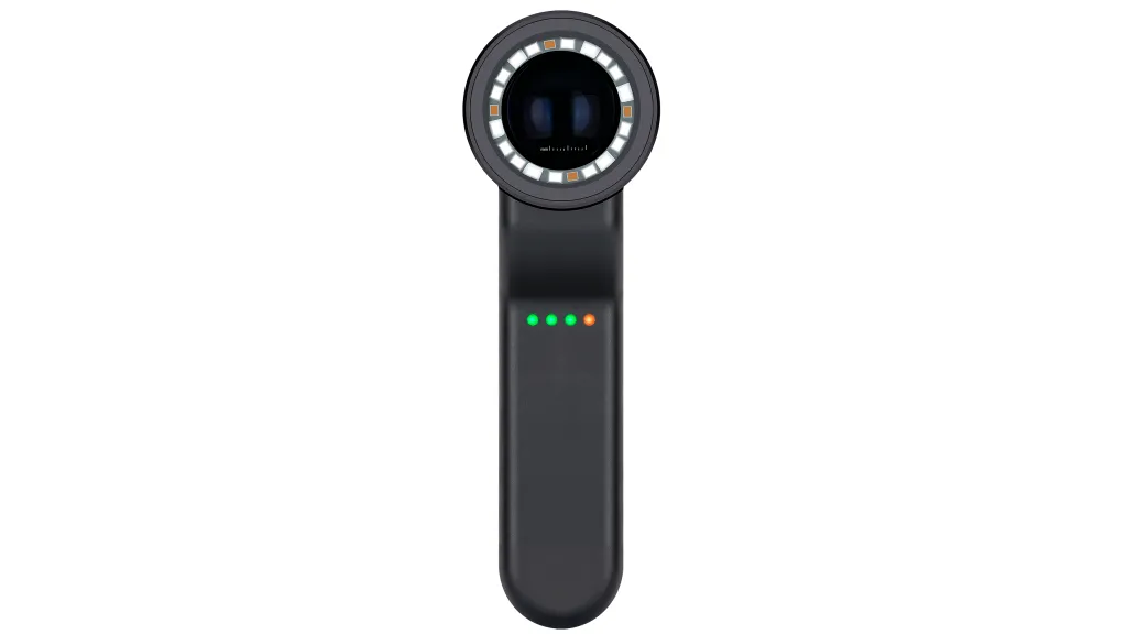
The IBOOLO DE-4100 and DE-3100 Dermatoscopy Optical Magnification Systems magnify skin lesions up to ten times through a combination of multiple glass lenses. Both dermatoscopes utilize the principle of polarized light, which enhances the collection of transmitted light by adjusting the direction of light polarization to reduce the interference of reflected light from the skin surface. This technology allows the dermatoscope to penetrate deep into the skin tissue and observe the structure and characteristics of the epidermis and dermal papillary layers.
Dermoscopic Features of Basal Cell Carcinoma
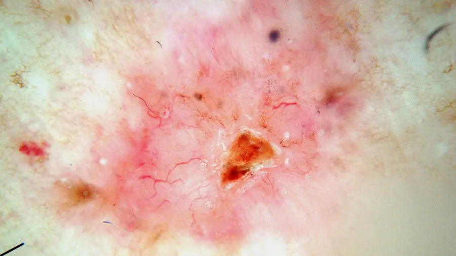
Dermoscopy can acutely capture subtle color distinctions and changes in skin lesions, and therefore, dermoscopy has significant advantages in the diagnosis of skin diseases such as pigmented nevi, malignant melanoma and basal cell carcinoma. The following is an example of dermoscopic examination of basal cell carcinoma.
Dermoscopic features of basal cell carcinoma mainly include the following aspects:
Large blue-gray ovoid nests: this is one of the classic dermoscopic features of pigmented basal cell carcinoma, which is manifested by the appearance of multiple blue-gray ovoid structures of varying sizes in the lesion area.
Multiple blue-gray spherules: these spherules are usually distributed in a non-aggregated fashion and are another classic dermoscopic feature of basal cell carcinoma.
Spoke-like areas: Spoke-like areas appear as structures radiating from the center of the lesion to the periphery.
Importance of Dermoscopy Examination for Early Detection of Basal Cell Carcinoma
Basal cell carcinoma, a kind of malignant tumor originated from basal-like cells, is one of the most common skin malignant tumors, and its incidence rate is very high.BCC grows slowly but has the characteristic of local infiltrative growth, which is capable of destroying tissues and organs, and even threatens the patients’ lives in serious cases.
Dermoscopy plays a crucial role in the early detection of basal cell carcinoma. Dermoscopy examination can quickly capture many typical features of basal cell carcinoma, such as dendritic blood vessels, superficial short capillary dilatation, bleeding ulcers and so on. In addition, dermoscopy can also assess the infiltration depth and scope of basal cell carcinoma, which provides an important basis for the formulation of treatment plan.
IBOOLO Dermatoscope
IBOOLO has different series of dermatoscopes that offer different options for dermatoscopic examination. DE-200, DE-300 and DE-400 are pocket dermatoscopes that are compact and can be carried around. The DE-200, DE-300 and DE-400 are handheld dermatoscopes that can be used to view lesions directly through a window or to connect to a cell phone via a magnet. the DE-200, DE-300, and DE-400 are relatively inexpensive and have polarization, making them ideal for those who require only basic dermatoscopic functionality. the DE-3100 and DE-4100 are relatively inexpensive and have polarization, making them ideal for those who require only basic dermatoscopic functionality. The DE-3100 and DE-4100 are more expensive and have a variety of illumination sources and brightness adjustments, making them ideal for professionals.
Dermoscopy Procedure
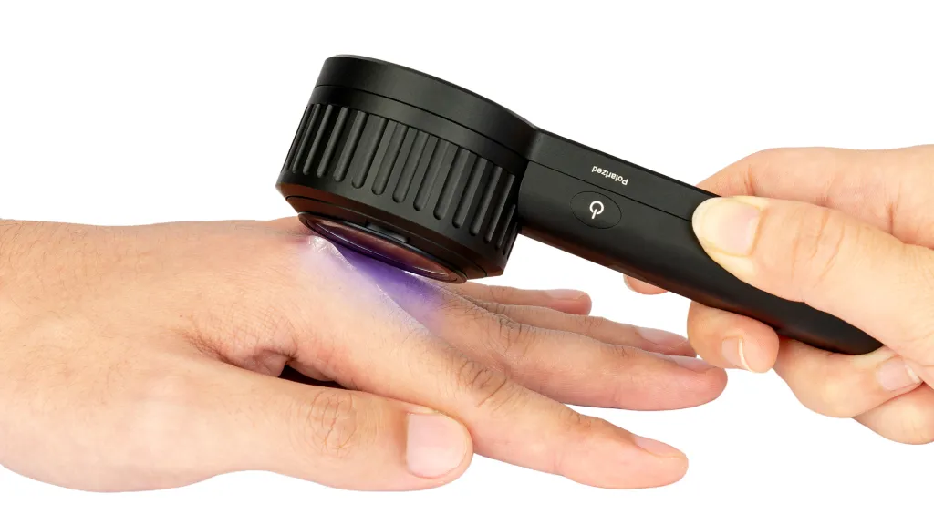
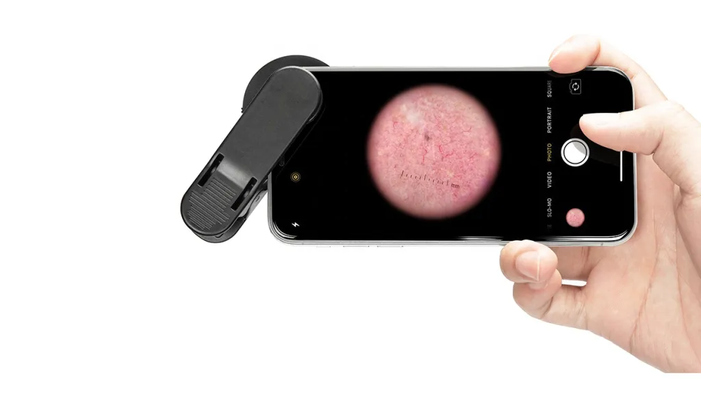
The following is the procedure for a dermoscopy from start to finish. First, the surface of the patient’s skin that needs to be examined needs to be cleaned to ensure that it is dry and free of oil and dirt. Next, the probe of the dermatoscope is attached to the skin surface that needs to be observed. Keep the distance between the probe and the skin, adjust the focus until the image is clear to start observation. You can connect the dermatoscope to your cell phone to save photos of skin lesions. The doctor can judge the disease based on the skin pigmentation and blood vessel changes in the dermatoscope window, and suggest the appropriate treatment plan for the patient.
Advantages of the Dermoscopy Procedure
The entire process of dermoscopy usually takes less than 10 minutes, making it a quick and easy procedure. It enables doctors to obtain detailed information about the skin surface in a short period of time. Moreover, dermoscopy is a non-invasive and non-invasive diagnostic method that does not cause any damage to the skin or any discomfort to the patient, making it easier for the patient to accept this method of examination.
Dermoscopy Examination and Specialized Training
Dermoscopy is only an examination tool to provide professionals with a good viewing field. The final result of the examination still largely depends on the experience as well as the knowledge of the doctor. Therefore, it is important for healthcare professionals to receive systematic training on the basic principles of dermoscopy, its operation and the dermoscopic features of common skin diseases. There is a wide variety of skin diseases and new diseases and variants are constantly emerging. Healthcare professionals need to continually update their knowledge base on the latest dermoscopic diagnostic techniques and research findings by attending seminars and training courses.
Conclusion
The key to dermatoscopy is the use of optical magnification and polarization modes to help the physician better visualize the lesions, and IBOOLO has a wide range of dermatoscopes to meet your different needs. For more information about IBOOLO dermatoscopes, please visit the product section of the website. Dermoscopy is valuable in diagnosing skin tumors, evaluating skin inflammation, detecting hair diseases, and identifying parasitic infections. It can help doctors determine the nature of skin lesions, such as benign or malignant, and provide guidance for surgical removal. IBOOLO dermatoscope can definitely provide to be your most reliable seeing instrument in the daily visits of dermatologists.
Dermoscopy Examination: A Comprehensive Guide to Skin Cancer Detection | IBOOLO
Discover how dermoscopy can help detect skin cancer early. Learn about IBOOLO dermatoscopes, their features, and benefits for dermatologists.
Dermoscopy Examination: Key Technology and Clinical Applications for Early Skin Cancer Detection
The incidence of skin cancer continues to rise annually, making early detection crucial for improving patient prognosis. Dermoscopy, as a non-invasive diagnostic tool, utilizes optical magnification and polarized light technology to help physicians observe both superficial and deep skin structures, capturing pigment and vascular changes that are invisible to the naked eye. This article analyzes the core technology of dermoscopy, its clinical value, examination procedures, and its important role in early skin cancer diagnosis, while exploring its applications across different age groups and skin conditions through real case studies.
Introduction and Core Technology
What is the Key Technology of Dermoscopy in the Early Detection of Skin Cancer? Experts Explain in Detail
Skin cancer has become an increasingly prominent health concern in recent years. With the rise in the incidence rate, early detection has emerged as a crucial factor in improving the prognosis of patients. Dermoscopy, also known as dermatoscopy or chemiluminescence microscopy, plays a pivotal role in this process.
The Principle of Dermoscopy in Detecting Skin Cancer
Dermoscopy enables doctors to observe the skin surface and subsurface structures in greater detail compared to the naked eye. It uses polarized light or immersion liquid to reduce the reflection of the skin surface, thus allowing for a clearer view of pigmented and vascular patterns within the skin. For example, in the case of melanoma, dermoscopy can detect characteristic features such as irregular pigment network, blue-white veil, and atypical vascular patterns. These features are often not visible to the unaided eye in the early stages of the disease.
Specific Technical Details
Magnification: Dermoscopes typically offer magnification ranging from 10x to 40x. This magnification helps in identifying subtle changes in skin lesions. For instance, small dots or globules within a mole, which may be indicative of melanoma, can be more easily detected under appropriate magnification.
Light Source: The use of polarized light is a key aspect. Polarized light penetrates the skin more effectively, reducing the interference from the stratum corneum. This results in a clearer visualization of the underlying structures. In some advanced dermoscopes, different wavelengths of light can be used to enhance the contrast of specific skin components, further improving diagnostic accuracy.
Clinical Value Compared to Regular Examinations
Why Do Dermatologists Recommend Dermoscopy Over Regular Physical Examinations?
When it comes to skin health, many people rely on regular physical examinations. However, dermatologists often recommend dermoscopy as a more effective diagnostic tool. Let's explore the reasons behind this preference.
Superior Visualization of Skin Structures
Enhanced Detail: Regular physical examinations rely solely on the naked eye. Dermoscopy, on the other hand, uses magnification and specialized lighting techniques. As mentioned before, dermoscopy can magnify the skin up to 40x, allowing dermatologists to see minute details such as individual pigment granules, small blood vessels, and subtle changes in the texture of the skin. For example, a small change in the pigmentation pattern of a mole that might be overlooked during a regular physical examination can be seen under dermoscopy.
Sub-surface Visualization: Dermoscopy can penetrate beneath the skin surface. Reducing the reflection from the stratum corneum enables doctors to view structures in the epidermis and upper dermis. This is particularly important as many skin cancers originate in these layers. In contrast, a regular physical examination can only provide information about the surface appearance of the skin.
Higher Diagnostic Accuracy
Early Detection of Skin Cancer: The ability to detect subtle changes in skin lesions at an early stage is crucial in the fight against skin cancer. Dermoscopy has been shown to significantly improve the accuracy of diagnosing melanoma, the most dangerous form of skin cancer. Studies have indicated that the use of dermoscopy can increase the diagnostic accuracy of melanoma by up to 30 40% compared to clinical examination alone. This early detection can lead to more effective treatment and better patient outcomes.
Differentiation of Benign and Malignant Lesions: Dermoscopy helps dermatologists distinguish between benign moles, seborrheic keratoses, and other non-cancerous skin growths and potentially malignant lesions. By analyzing pigmentation patterns, vascular structures, and the overall morphology of the lesion, doctors can make more accurate diagnoses. In a regular physical examination, it can be challenging to differentiate between some benign and malignant lesions based on visual inspection alone.
The Examination Process
How to Conduct a Dermoscopy Examination: A Complete Guide from Procedure to Result Interpretation
Dermoscopy is a non-invasive diagnostic technique that has become an essential part of dermatological practice. Understanding the entire process, from the moment you enter the examination room to the interpretation of the results, can help you better prepare and make sense of the information.
Procedure of Dermoscopy
Patient Preparation: First, the patient is asked to lie down or sit in a comfortable position. The area of the skin to be examined is then exposed. It is important to ensure that the skin is clean and free of any creams, lotions, or makeup, as these can interfere with the quality of the dermoscopy image.
Application of Immersion Liquid (Optional): In some cases, an immersion liquid such as mineral oil or alcohol-based gel is applied to the skin surface. This helps to reduce the reflection from the skin surface and provides a clearer view of the underlying structures. The liquid is spread evenly over the area of interest using a cotton swab or a similar applicator.
Using the Dermoscope: The dermatologist then holds the dermoscope close to the skin, usually at a right angle. The dermoscope is moved slowly across the skin, scanning the entire area of the lesion. The doctor may adjust the magnification and focus of the dermoscope as needed to get a detailed view. During this process, the doctor looks for various features such as pigmentation patterns, vascular structures, and the overall shape and border of the lesion.
Image Capture (If Available): Many modern dermoscopes are equipped with cameras. If so, the doctor will capture images of the lesion from different angles. These images can be used for further analysis, comparison with previous examinations, or for sharing with other specialists.
Result Interpretation
Pigmentation Patterns: A regular pigment network may indicate a benign mole, while an irregular pigment network could be a sign of melanoma. The presence of blue-white areas may also be associated with melanoma.
Vascular Patterns: Atypical vascular patterns, such as dotted, linear-irregular, or hairpin-shaped vessels, are often seen in melanoma or other skin malignancies. In contrast, benign lesions usually have more regular vascular patterns.
Border and Shape: Lesions with an irregular border and asymmetrical shape are more likely to be malignant. Benign moles typically have smooth, well-defined borders and a symmetrical shape.
Detection Capabilities
What Skin Problems Can Dermoscopy Detect?
Dermoscopy is a powerful tool that can reveal a wide range of skin problems. By providing a detailed view of the skin surface and subsurface, it helps dermatologists make accurate diagnoses.
Pigmentation Related Skin Problems
Melanoma: As previously mentioned, dermoscopy is highly effective in detecting melanoma. It can identify characteristic features such as the ABCDE criteria (asymmetry, irregular border, colour variegation, diameter larger than 6mm, and evolving changes) at an early stage. The presence of an irregular pigment network, blue-white veil, and atypical vascular patterns are strong indicators of melanoma.
Solar Lentigines: These are also known as age spots or liver spots. Dermoscopy can show a characteristic pattern of small, round, or oval-shaped pigmented areas. The pigmentation is usually uniform, and there are no signs of malignancy.
Melasma: This is a common skin condition characterized by brown or grey-brown patches on the face. Dermoscopy can help distinguish melasma from other pigmentary disorders. It may show a network of fine brown lines, which are due to increased pigmentation in the epidermis and dermis.
Vascular Related Skin Problems
Telangiectasia: These are small, dilated blood vessels that are visible on the skin surface. Dermoscopy can clearly show the shape and distribution of these vessels. They often appear as fine, red lines or dots, and can be associated with various skin conditions such as rosacea.
Hemangiomas: Dermoscopy can help identify different types of hemangiomas. For example, infantile hemangiomas may show a characteristic lobular pattern with red-purple nodules, while cherry hemangiomas appear as small, round, bright red papules.
Other Skin Problems
Seborrheic Keratoses: These are common, benign skin growths. Dermoscopy can show a characteristic pattern of keratin-filled cysts, also known as pseudo-horn cysts, and a cerebriform (brain-like) surface texture.
Actinic Keratoses: These are precancerous lesions. Dermoscopy can detect features such as scale-crust, follicular plugging, and a pink-red background with telangiectasia, which is characteristic of actinic keratoses.
What Can Dermoscopy Detect?
Dermoscopy can help detect various common skin conditions. Here are several typical cases:
Case 1: Basal Cell Carcinoma
Symptoms: The patient presented with a slightly raised red patch on the face, accompanied by mild itching.
Dermoscopic Features:
Blue-gray ovoid nest structures
Arborizing telangiectasia (branching blood vessels)
Slight surface scaling
Diagnostic Outcome: Through dermoscopic examination, the doctor made a preliminary diagnosis of basal cell carcinoma, which was subsequently confirmed by biopsy.
Case 2: Melanoma
Symptoms: The patient noticed recent changes in the colour and shape of a mole on their back.
Dermoscopic Features:
Uneven pigment distribution with pseudopod-like extensions
Blurred borders with varying colour intensity
Diagnostic Outcome: Dermoscopy indicated possible malignant melanoma, later confirmed as early-stage melanoma through biopsy.
Case 3: Seborrheic Keratosis
Symptoms: The patient developed multiple brown patches on the arm with rough surfaces.
Dermoscopic Features:
Well-defined "brain-like" structures
Visible keratin plugs on the surface
Diagnostic Outcome: Dermoscopic examination confirmed benign seborrheic keratosis requiring no further treatment.
Why Are These Cases Important?
These cases demonstrate the crucial role dermoscopy plays in distinguishing between benign and malignant lesions, helping both doctors and patients make more informed decisions.
Professional vs Home Use
What is the Difference Between Home and Professional Dermoscopy?
Home Dermoscopy:
Portable design suitable for routine self-examination
Relatively simple functionality, primarily for preliminary observation
Professional Dermoscopy:
High-resolution imaging with multiple light source modes
Requires professional physician operation and result interpretation
How to Choose the Right Option for You?
1. Home dermoscopy is suitable for:
People who wish to monitor skin changes regularly
High-risk individuals with a family history of skin cancer
Those who need preliminary screening for skin issues
2. Professional dermoscopy is suitable for:
People who have already identified suspicious skin lesions
Those requiring precise diagnosis and treatment recommendations
Individuals seeking high-quality images through professional equipment
Why is Professional Dermoscopy More Reliable?
Greater accuracy: Professional equipment captures more details
Expert interpretation: Physicians can make accurate judgments based on clinical experience
Comprehensive assessment: Supports diagnosis and tracking of various skin conditions
What Should You Ask After a Dermoscopy Examination?
1. Is my skin lesion benign or malignant?
Understand the nature of the lesion and determine whether further treatment is necessary
2. Is a biopsy or additional testing needed?
Confirm whether dermoscopy is sufficient for diagnosis or if supplementary tests are required
3. What is the development trend of the lesion?
Understand if the lesion is stable, progressing, or regressing to establish a subsequent monitoring plan
4. What treatment options are available?
Learn about the advantages and disadvantages of surgical, medicinal, or other treatment methods based on the nature of the lesion
5. How can I prevent similar issues?
Obtain skin health advice such as sun protection, skincare, and regular check-ups
How to Read Your Dermoscopy Report?
Lesion Description: Note characteristics such as colour, shape, and borders
Diagnostic opinion: Understand the physician's preliminary judgment and recommendations
Image records: Compare lesion images to observe trends in changes
Why Are These Questions Important?
These questions help patients comprehensively understand their skin health status and actively participate in treatment decisions.
Cost Considerations
Does the Cost of Dermoscopy Justify the Investment in Skin Health?
The cost of medical procedures is always a concern for patients. When it comes to dermoscopy, understanding the cost-effectiveness of skin health is important.
Breakdown of Dermoscopy Costs
Equipment Cost: Dermatologists need to invest in high-quality dermoscopes. These can range in price from a few hundred dollars for basic models to several thousand dollars for more advanced, digital dermoscopes with imaging capabilities. The cost of the equipment is factored into the overall cost of the procedure for the patient.
Professional Fee: The dermatologist's time and expertise also contribute to the cost. Performing a dermoscopy examination requires training and experience to accurately interpret the findings. The professional fee varies depending on the location, the reputation of the dermatologist, and the complexity of the case.
Additional Costs (If Any): In some cases, there may be additional costs such as the use of immersion liquid, which is relatively inexpensive, or if images are printed or stored for future reference, there may be a small charge for the associated materials and data storage.
Value in Terms of Skin Health
Early Detection of Skin Cancer: As we know, early detection of skin cancer through dermoscopy can save lives. The cost of treating skin cancer at an early stage is often significantly lower than if the cancer has advanced. For example, a melanoma detected early may require only a simple excision, while advanced melanoma may need more extensive surgery, chemotherapy, or immunotherapy, which are far more expensive.
Peace of Mind: For patients who are concerned about their skin health, dermoscopy can provide peace of mind. Knowing that a potential skin problem has been thoroughly evaluated and accurately diagnosed can reduce anxiety. This psychological benefit is also an important aspect of the value of dermoscopy.
Preventive Care: Dermoscopy can also be used as a preventive tool. By detecting precancerous lesions such as actinic keratoses early, appropriate treatment can be initiated to prevent them from developing into invasive skin cancer. This proactive approach can save both health and money in the long run.
What is AI-assisted dermoscopy?
AI-assisted dermoscopy uses artificial intelligence algorithms to analyze dermoscopic images, helping doctors diagnose skin lesions more quickly and accurately.
How Does AI Improve Dermoscopy?
1. Enhanced diagnostic efficiency: AI can analyze images within seconds, providing preliminary diagnostic opinions
2. Reduced human error: Through big data training, AI can identify subtle lesion characteristics
3. Support for telemedicine: Patients can receive professional diagnostic advice by uploading images
Why is AI the Future of Dermoscopy?
Precision: AI can identify lesion features that are difficult to detect with the naked eye
Accessibility: Makes high-quality skin examinations available to more people
Innovation: Drives continuous advancement in dermatological diagnostic technology
Why is Age a Factor in Dermoscopy?
Different age groups face different skin health risks, resulting in varying needs for dermoscopic examinations.
How Often Should You Get a Dermoscopy?
Ages 20-30:
Examination every 2-3 years, focusing on newly appearing moles or patches
Ages 30-50:
Annual examination, especially for high-risk individuals (such as those with prolonged sun exposure)
Ages 50+:
Examination every 6-12 months, focusing on early signs of skin cancer
What Are the Risk Factors to Consider?
Family history: People with a family history of skin cancer should have more frequent examinations
Sun exposure habits: People with prolonged sun exposure need regular monitoring
Skin type: People with fair skin or skin prone to sunburn have a higher risk
Why is Regular Dermoscopy Important?
Regular dermoscopic examinations can help detect skin issues early, significantly improving treatment effectiveness and quality of life.
Dermoscopy, with its unique key technologies, provides a powerful tool for the early detection of skin cancer. Leveraging magnification and advanced light sources, allows doctors to identify potential skin cancer cases at a stage when treatment is often more effective.
Dermoscopy examination significantly enhances the precision of skin lesion observation through optical magnification and polarized light technology, showing exceptional performance in early skin cancer diagnosis. Its core technology includes high-power magnification and polarized light sources, which display pigment distribution and vascular structures, helping physicians distinguish between benign and malignant lesions. Compared to conventional physical examinations, dermoscopy offers higher diagnostic accuracy, enabling early detection of melanoma, basal cell carcinoma, and other skin cancers. The examination process is simple and non-invasive, suitable for people of all age groups, especially high-risk populations. Looking forward, the introduction of AI-assisted diagnostic technology will further enhance the efficiency and accuracy of dermoscopy examinations, providing more comprehensive support for skin health management.


