


Article
Dermoscopy Applications in the Diagnosis of Psoriasis
Dermoscopy of Psoriasis What is Psoriasis? Psoriasis is a long term skin disease which can grow anywhere but most commonly appear on the elbows, knees, scalp and trunk. Psoriasis is characterized by rash with itchy, scaly patches. It is a painful chronic disease and with no cure, which means that symptoms appear unexpectedly and may…
Dermoscopy of Psoriasis: A Comprehensive Overview - IBOOLO
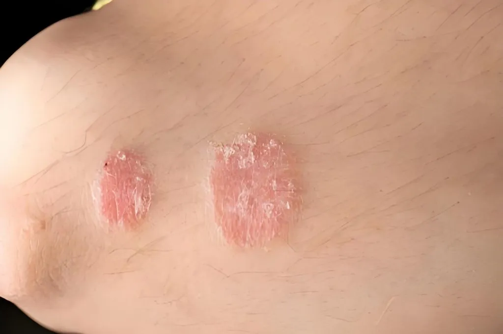
Dermoscopy of psoriasis enables non-invasive diagnosis through hallmark patterns: regularly distributed red-dotted vessels, white scales, and a light-red background.
Dermoscopy of Psoriasis: A Key Tool for Diagnosis and Management
The dermoscopy of psoriasis has transformed dermatological diagnostics, offering a non-invasive way to visualize skin structures beyond the naked eye. Known as dermatoscopy, this technique uses optical magnification and polarized light to reveal detailed patterns, such as vascular networks and scales, critical for diagnosing psoriasis. This article provides a comprehensive guide to the dermoscopy of psoriasis, exploring its hallmark features, applications across psoriasis subtypes, and its role in severity assessment and treatment monitoring, tailored for medical professionals, patients, and those curious about advanced skin imaging.
Why Dermoscopy Matters for Psoriasis
Enhancing Diagnostic Precision
The dermoscopy of psoriasis is a game-changer for identifying this chronic inflammatory condition, which affects millions globally with erythematous plaques and silvery scales. Unlike invasive biopsies, dermoscopy offers a patient-friendly approach, magnifying epidermal and dermal details like vascular patterns and scale morphology. This enhances diagnostic accuracy, especially for atypical cases, making dermoscopy of psoriasis a vital tool in clinical practice.
Broad Clinical Applications
Beyond diagnosis, the dermoscopy of psoriasis aids in differentiating it from similar conditions, assessing disease severity, and monitoring treatment outcomes. Its non-invasive nature makes it ideal for repeated use, providing insights into disease progression and therapeutic efficacy.
Dermoscopic Features of Psoriasis Vulgaris
Vascular Patterns
The dermoscopy of psoriasis vulgaris, the most common subtype, reveals distinct vascular features:
- Regular Red-Dotted Vessels: Symmetrically arranged pinpoint capillaries, seen in 96–100% of cases, correspond to dilated vessels in dermal papillae. Their uniform distribution has 100% specificity for psoriasis.
- Ring/Hairpin-Like Vessels: Less common (3.6–44.1%), these coiled or lacelike capillaries offer high diagnostic specificity (91.9–100%).
Scale Patterns
Scales are a hallmark of psoriasis vulgaris under dermoscopy:
- White Scales: Found in 64.7–88.3% of patients, these reflect orthokeratosis or parakeratosis, with 83.8% specificity. They are diffuse in older lesions and patchy in newer ones.
- Yellow Scales: Seen in 2–25.2% of cases, often in older patients, due to slower skin turnover.
Additional Features
Other dermoscopic signs include:
- Light Red Background: Common in psoriasis, with 71–78% sensitivity, reflecting epidermal thinning and inflammation.
- Hemorrhagic Dots: Present in ~30% of cases, often on lower limbs, linked to scratching or venous stasis.
The dermoscopy of psoriasis vulgaris typically shows regular dotted vessels, white scales, and a light red background, offering 88% specificity and 84.9% sensitivity for diagnosis.
Dermoscopy Across Psoriasis Subtypes
Nail Psoriasis
The dermoscopy of psoriasis in nails enhances visualization of features like pitting, subungual hyperkeratosis, and splinter hemorrhages. Unique signs, such as dilated hyponychial capillaries and the pseudofiber sign, are often only visible through dermoscopy, improving diagnostic accuracy.
Palmoplantar Psoriasis
In palmoplantar psoriasis, dermoscopy reveals diffuse white scales and less frequent dotted vessels due to thicker skin. Beaded vessel patterns along skin furrows are a key diagnostic clue.
Scalp Psoriasis
The dermoscopy of psoriasis on the scalp shows red-dotted vessels, twisted red loops, and thick white scales. Signet ring vessels and hidden hair further support diagnosis.
Other Variants
- Erythrodermic Psoriasis: Features regular dotted vessels and white scales on a light red background.
- Inverse Psoriasis: Shows dotted vessels on a reddish background with minimal scales.
- Guttate Psoriasis: Displays dotted vessels and white scales, less pronounced due to smaller lesions.
- Pustular Psoriasis: Reveals milky globules (sterile pustules) and dotted vessels on a reddish background.
Differentiating Psoriasis with Dermoscopy
Key Differential Diagnoses
The dermoscopy of psoriasis aids in distinguishing it from conditions with similar presentations:
- Lichen Planus: Shows linear vessels and Wickham striae, unlike psoriasis’ dotted vessels.
- Pityriasis Rosea: Features patchy dotted vessels and fine peripheral scales.
- Mycosis Fungoides: Displays spermatozoa-like vessels and fine geometric scales.
- Seborrheic Dermatitis (Scalp): Shows arborizing red lines and greasy scales, contrasting with psoriasis’ dotted vessels.
- Onychomycosis (Nails): Exhibits spikes and ruin patterns, unlike psoriasis’ pitting and hemorrhages.
These distinctions highlight the dermoscopy of psoriasis as a critical tool for accurate differentiation.
Assessing Severity and Treatment Response
Evaluating Disease Severity
The dermoscopy of psoriasis supports severity assessment through tools like the Vascular Psoriasis Area Severity Index (VPASI). Nail dermoscopy, showing features like plate thickening, correlates with systemic inflammation and severity scores like NAPSI.
Monitoring Treatment Efficacy
Changes in dermoscopic features, such as reduced vessel density or scale resolution, indicate treatment success. In nail psoriasis, decreased visible capillaries and improved scaling reflect positive responses to therapy.
Predicting Outcomes and Recurrence
Treatment Outcome Prediction
The dermoscopy of psoriasis can predict therapeutic responses. Globular vessels may indicate treatment resistance, while dotted vessels suggest better outcomes with topical therapies. Hemorrhagic dots may predict favorable responses to biologics.
Monitoring Recurrence
Persistent dotted vessels after clinical remission signal a higher recurrence risk, introducing the concept of “dermoscopic healing.” Their reappearance may predict relapse post-treatment, aiding proactive management.
Limitations of Dermoscopy in Psoriasis
Despite its strengths, the dermoscopy of psoriasis faces challenges:
- Limited Research: Current studies are few, requiring larger, multicenter trials.
- Inconsistent Terminology: Varied descriptions of dermoscopic features, especially in nail psoriasis, need standardization.
These limitations underscore the need for ongoing research to refine diagnostic criteria.
The Power of Dermoscopy in Psoriasis Care
The dermoscopy of psoriasis is a non-invasive, cost-effective tool revolutionizing the diagnosis, differentiation, and management of this chronic condition. By revealing specific patterns like dotted vessels and white scales, it enhances clinical precision across psoriasis subtypes. While limitations exist, its potential in severity assessment, treatment monitoring, and outcome prediction makes it a cornerstone of modern dermatology. Always consult a dermatologist to interpret dermoscopy findings, ensuring comprehensive care for psoriasis management.
Recommended reading
Wholesale Dermascope Vs Dermatoscopes Products Supply by Company in China - IBOOLO
We are a leading China-based dermascope vs dermatoscope products supply capable of providing wholesale lamps designed around our clients' specific needs. Our flexible products supply process ensures tailored solutions.
portable handheld dermatoscope precise skin lmaging - IBOOLO
IBOOLO DE-4100 handheld dermatoscope enables detailed magnified skin analysis anywhere with its lightweight, ergonomic design. customizable polarized lighting captures clear subsurface skin views.
Custom Case for iPhone suppliers & factories – IBOOLO
IBOOLO is a Custom Case for iPhone suppliers & factories. Information for Case for iPhone: Case for iPhone – Compatible with Dermatoscope...
Dermoscopy of Psoriasis
What is Psoriasis?
Psoriasis is a long term skin disease which can grow anywhere but most commonly appear on the elbows, knees, scalp and trunk. Psoriasis is characterized by rash with itchy, scaly patches.
It is a painful chronic disease and with no cure, which means that symptoms appear unexpectedly and may go throughout the whole life. It trends to go through a cycle where it flares for a few weeks or months and then subsides for a while.The condition interferes with sleep and concentration and also varies in severity.
How does dermoscopy detect and diagnose the types of psoriasis skin disease?
Dermoscopy, also known as dermatoscopy, is a non-invasive tool aiding dermatologists to clearly observe skin lesions which are invisible to the naked eye. To help to diagnose psoriasis, dermoscopy can reveal specific patterns and features of it. The characteristics of dermoscopy of psoriasis as below:
• Dotted vessels: Dotted vessels are the most common dermoscopic features inspected in psoriasis. They show as tiny dots within the psoriatic plaques. If dermscope detect any other morphologic type of vessels, then it can exclude the psoriasis diagnosis.
• Red globules: Sometimes called dots or balls, they correspond to vertically arranged rings of blood vessels within slender dermal papillae. They may differ in diameter, but are usually of similar size within a given lesion.
• Uniform distribution: These vessels at the lesion site showing as symmetrical and uniform distribution is a landmark of psoriatic plaques.
• Removing scales: Removing scales can display tiny red blood drops and reveal the characteristic vascular pattern of psoriasis, called as the dermoscopic” Auspitz” sign.
• Red globular rings: Although red globular rings are rare, but for psoriasis, it is a high specification that circles or rings of red balls present irregularly.
During treatment, dermalogists also can monitor the processing or the transformation of psoriatic plaques with the aid of dermoscope. Thus dermoscopy of psoriasis can provide extra morphological information which may be very useful for early examination of relapse.
How distinguish between psoriasis and eczema under dermoscope?
When using a dermoscope to distinguish between psoriasis and eczema, there are some main characteristics for consideration as below:
Color
Variation: Under dermoscope, psoriatic plaques exhibit a uniform salmon pink color.
Eczema: Eczematous lesions tend to have more various colors, red, yellow, blue or brown are included. These color may change according to the stage of inflammation.
Vascular Patterns:
Psoriasis : Under dermoscopy, psoriasis lesions usually present glomerular blood vessels or regular punctal blood vessel, which is also called “strawberry pattern” ). Most of these vessels are evenly distributed within the lesion part.
Eczema: Eczematous lesions often exhibit more sparse and irregular blood vessels. often showing a linear or linear irregular pattern. These vessels can be less obvious than in psoriasis.
Micro-Hemorrhages:
Psoriasis: Look for pinpoint red dots (micro-hemorrhages) within psoriatic plaques. These dots represent dilated capillaries and are characteristic of psoriasis. Look for tiny red spots (micro-bleeds) in patches of psoriasis. These dots means angiotelectasis, a characteristic of psoriasis.
Eczema: In eczema lesions, micro-hemorrhages are infrequent.
Scale and Crusts:
Psoriasis:Under dermoscope, psoriatic patches usually have silver and white scales with shiny looks.These scales characters thickness and adhesion.
Eczema: Eczematous lesions may appear tiny white scales, and they are less obvious than psoriasis. In addition, eczema lesions may scab due to exudation or scratches.
Distribution and Symmetry:
Psoriasis:Psoriasis patches are usually symmetrically distributed on the surface of the extensor muscles ( knees, elbows, scalp, lower back).
Eczema: Eczema lesions can occur in any part of the body and they are asymmetrical. They may be more common in curved areas (behind the knee,inside the elbow).
There are many key features to distinguish between psoriasis and eczema. Dermoscopy of psoriasis and eczema both can help to enhance the visual field for these skin conditions more closely. So that the dermatologist can make a accurate diagnosis combining dermoscope with clinical experience.
Is dermoscope the main tool for psoriasis diagnosis?
Yes, dermoscope is one of the valuable main tool for psoriasis diagnosis. Dermoscope plays a important role in the process of diagnosing psoriasis. But it is not the sole device for diagnosing psoriasis. There are other devices for helping to diagnose psoriasis more accurate and comprehensive, such as Wood’s Lamp Examination, Laboratory Tests, and Psoriasis Area and Severity Index (PASI).
Other Tools:
Wood’s Lamp Examination: Wood’s lamp can highlight psoriatic plaques by ultra violet examination because of increased fluorescence.
Laboratory Tests: Blood tests (such as C-reactive protein and erythrocyte sedimentation rate) may provide supporting evidence.Serum markers for autoimmune activity and inflammation are included.
Psoriasis Area and Severity Index (PASI): Based on lesion characteristics, such as the degree of skin severity, the intensity of erythema, scale, and thickness, the PASI assesses the severity of psoriasis.
Additionally, Dermatologists evaluate the patient’s skin by their history, clinical symptoms. Even when it is necessary, a skin biopsy is need to be performed.
While dermoscopy supplies clearly wide visual, a comprehensive approach and other tools are needed to ensures accurate psoriasis diagnosis.
What is the clinical value of dermoscopy in psoriasis?
Dermoscopy a noninvasive device contributing to important clinical value in the evaluation and management of psoriasis. There are some key clinical values for dermoscopy of psoriasis as below:
Psoriasis diagnosis
Identification of Psoriasis: Dermoscopy helps to identify typical features of psoriasis, like regular punctate blood vessels within the erythema plaques.
Timely diagnosis: Dermoscope aids dermologists to diagnose and interfere in timely, and improve the treatment outcome of patients.
Monitoring Disease Progression:
Objective Assessment: Dermoscopy is used to monitor changes in psoriasis over time, and it provides an objective way to assess the severity of psoriasis.
Quantifying Lesions: With using dermoscope, dermatologists can measure the extent of involvement, scaling and erythema of psoriasis.
Treatment Feedback: Dermoscope can monitor precisely the feedback to treatment, even including good fedback, bad feedback and side effect.
Reducing Biopsy Need:
Noninvasive Approach: Dermoscopy greatly reduce the unnecessary biopsies for skin.
Avoiding Invasiveness: Using a dermoscope to examine skin is a invasive and painless process which still can get an accurate diagnosis.
Guiding Treatment Decisions: Targeted Therapies: Under dermoscopy, clinician can choose reasonable treatment therapeutic scheme. Dermoscope is a guider for treatment decisions.
The findings of dermoscopy provide complement information for clinical evaluation. So that clinician can make more proper treatment decisions for psoriasis. Deroscopy of psoriasis is a great significance in examination and management of psoriasis.


