


Article
Dermatoscopes for Sale
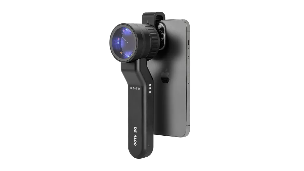
As a non-invasive diagnostic tool, handheld dermatoscopes typically offer a magnification of around 10 times. Dermatoscopes are commonly equipped with built-in light sources to ensure adequate and even illumination during the examination process. The IBOOLO Dermatoscopes DE-3100 and DE-4100 series are USB ports that allow users to charge the dermatoscope from common charging devices such as…
As a non-invasive diagnostic tool, handheld dermatoscopes typically offer a magnification of around 10 times. Dermatoscopes are commonly equipped with built-in light sources to ensure adequate and even illumination during the examination process. The IBOOLO Dermatoscopes DE-3100 and DE-4100 series are USB ports that allow users to charge the dermatoscope from common charging devices such as computers and rechargeable batteries without worrying about battery depletion. Moreover, both series of dermatoscopes have a polarization mode that eliminates the need for the user to reduce the scattering and reflection of light through the use of immersion liquids.
Imaging Requirements
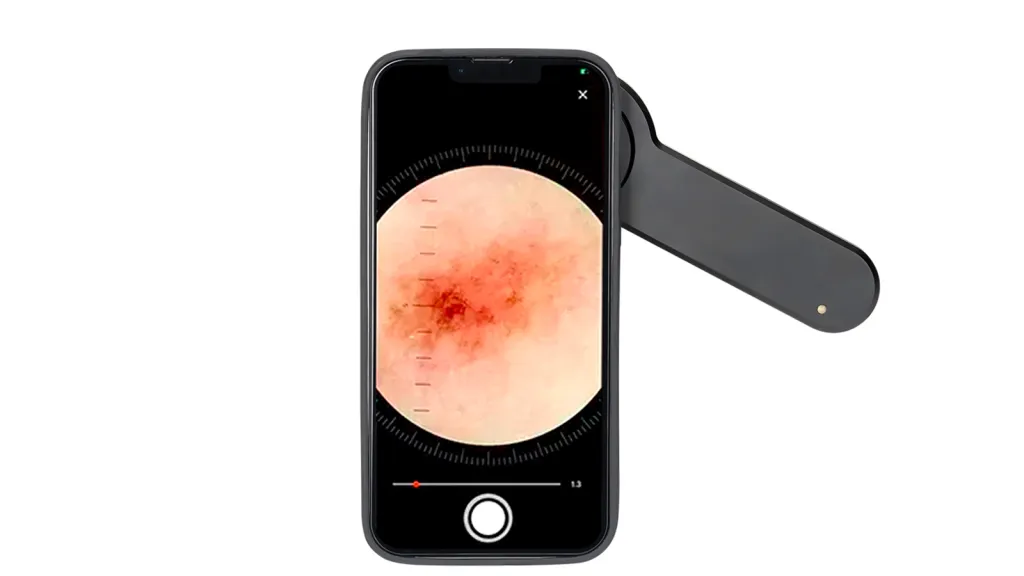
Handheld dermatoscopes are usually connected to a camera through a specific interface or adapter in order to transfer the observed skin images to the camera for recording and analysis. IBOOLO dermatoscopes can be connected to a camera or a cell phone through IBOOLO’s customized magnetic ring interface. This is a convenient and efficient way for the user to easily store and compare the images captured by the dermatoscope.
A high quality lens should have high optical resolution and low aberration, good light transmission and anti-reflection properties. High-quality sensors should have a high pixel count, wide dynamic range and low noise level.
Storage and transmission capabilities are equally important components of an imaging system. Adequate storage capacity and stable transmission capabilities allow for rapid transfer of image data to a computer or other device for further processing and analysis.
Smartphones and other personal mobile devices
Smartphones and other personal mobile devices offer unprecedented convenience with their intuitive interface and powerful features. Nowadays, according to most of the cell phones on the market are designed to be drop-proof, and are also paired with fingerprint or facial recognition, which is very sturdy and secure. The most important thing is that, whether it is social media, online shopping, or professional tool software, users can find the required applications in the app store, and easily download and install them on the device.IBOOLO Skin Dermoscopy can be connected to the cell phone to use, and by utilizing the powerful functions of the cell phone, the Skin Mirror can also take on a different look.
Available options for dermatoscopes
The combination of a smartphone adapter and a handheld device allows for a clear view of the fine structure of the skin by utilizing the existing high-resolution camera and powerful processing capabilities of a smartphone in combination with dermoscopic magnification, illumination and color correction.
A stand-alone dermatoscope camera with SD card is an ideal choice for users who require a higher degree of professionalism and independence. This device is usually equipped with a high-quality camera, a built-in light source and a specialized dermoscopic lens that provides a higher level of image quality and diagnostic capability than a smartphone adapter.
Considerations when choosing a dermatoscope
When choosing a suitable dermatoscope, we should first consider portability. If you have to use the dermatoscope frequently in your daily life or to examine lesions on the go, it is advisable to purchase a handheld dermatoscope, and the IBOOLO Handheld Dermatoscope DE-3100 can even be easily tucked into the pocket of your daily clothes. Secondly, the magnification of dermatoscopes also varies, with handheld dermatoscopes usually having a magnification of about 10x and digital dermatoscopes being able to reach dozens or even hundreds of times. However, digital dermatoscopes are expensive, so you need to consider the price when choosing the right one.
Dermatoscope Purchase Channel
The purchase channels of dermatoscopes can be simply categorized into online and offline. Online channels are mainly the major dermatoscope official websites and distributors of different shopping platforms, and IBOOLO dermatoscopes are mainly purchased through the IBOOlO official website. Some large dermatoscope brands may cooperate with major skin hospitals, where the dermatology department makes a unified purchase and then distributes it to doctors.
The difference between dermatoscopic examination and dermatoscope
The dermatoscope employs optical magnification and image enhancement technologies to clearly reveal details of the skin surface, such as texture, color variations, and vascular patterns. This process is known as dermatoscopic examination, which is a non-invasive and rapid method of skin inspection.
The dermatoscope, also referred to as the skin surface translucent microscope, is an instrument based on optical principles used for observing skin pigmentary disorders.
In summary, dermatoscopic examination is a specific application of the dermatoscope, which involves the process of inspecting the skin through the dermatoscope, while the dermatoscope itself serves as the tool or device utilized for such examination.
Accuracy of dermoscopy
First of all, we have to understand that dermoscopy is just a testing tool that clarifies and magnifies the skin lesions in the area to be observed. But the results of the examination this is judged by the doctor, that is to say, the doctor’s personal experience and subjective judgment may mislead the patient. Secondly, the accuracy of dermatoscopy is affected by its performance. High-quality and high-precision dermatoscopy equipment can provide clearer and more accurate images, thus improving the accuracy of diagnosis. The condition of the patient’s skin can also affect the results of dermoscopy. For example, uneven pigmentation or a strong epidermal inflammatory reaction may interfere with the doctor’s judgment, thus affecting the accuracy of the diagnosis.
Can dermoscopy detect cancer?
Dermoscopy can assist in the detection of cancer, especially skin cancer. Dermoscopy can help doctors more accurately identify abnormal changes in the skin, such as pigmented and non-pigmented lesions, which can aid in the diagnosis of malignant melanoma, basal cell carcinoma, squamous carcinoma and other skin tumors. The results of dermoscopy can provide important reference information for doctors and assist them in formulating treatment plans. If the results of dermoscopy show that there is a possibility of cancer, doctors will recommend further confirmatory tests such as skin pathology biopsy.
How to choose a dermatoscope
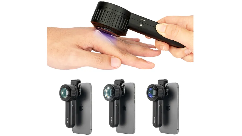
If you intend to purchase a dermatoscope that best suits your needs, you must thoroughly consider what your paramount requirements are. The IBOOLO DE-3100 and DE-4100 are highly specialized dermatoscopes equipped with polarized, non-polarized, and amber light functions, enabling doctors to observe skin lesions from multiple angles and thereby make more professional judgments. On the other hand, the IBOOLO DE-400 and DE-500 are simplified versions of dermatoscopes that require pairing with digital devices such as mobile phones to display images. This series falls under IBOOLO’s entry-level dermatoscope range, making it an ideal and cost-effective choice for non-professionals who wish to explore skin lesions on their own.
Recommended reading
Comparing Top Dermatoscopes to Dermatoscopio Comprar1. IBOOLO DL1 Wireless Dermatoscope
The DL1 from IBOOLO is an excellent choice if you re looking to dermatoscopio comprar a high-quality device at an affordable price. With premium optics, bright LED light, and a portable wireless design, it s great for both professionals and personal use. It can also connect to smartphones for image capture.
The Convenience of Handheld and Smartphone Dermatoscopes
Regular skin self-examinations are crucial for detecting suspicious moles or lesions early, when skin cancers are most treatable. However, thoroughly inspecting hard-to-see areas like the scalp, back, and between toes can be challenging. This is where compact handheld dermatoscopes and smartphone dermatoscopes provide a convenient solution for at-home skin screening.
Who Can perform Dermoscopy?
The powerful magnification and lighting capabilities dermatoscopes offer provide beneficial visual data for everyone to understand the current state of their skin and track changes over time. However, specialized medical training is typically required to analyze dermoscopy images and determine if biopsies or treatment are necessary. Dermatologists have this expertise.


