Article
Can a dermoscopy detect melanoma
Melanoma is a dangerous skin cancer, which threatens the lives of people all over the world.It starts from melanocytes. Melanocytes is a type of pigment-producing cell and can spread rapidly without early treatment. Though melanoma is very serious, it always be ignored beacuse of its inapparent sympyoms. These symptoms make many cases difficult to dectect….
Melanoma is a dangerous skin cancer, which threatens the lives of people all over the world.It starts from melanocytes. Melanocytes is a type of pigment-producing cell and can spread rapidly without early treatment. Though melanoma is very serious, it always be ignored beacuse of its inapparent sympyoms. These symptoms make many cases difficult to dectect. Therefore,it is important for us to take regular skin examinations. However, doctors feel difficult to distinguish whether a scar is harmful or not just by visual observation. That’s why we need dermoscopes ,the easy-to-work handheld device. It can magnify and illuminate layers of skin to reveal hidden details of moles. Dermoscope will help doctors to improve the accuracy of detecting melanoma. It also can reduce needless surgery. We are glad to tell you how dermoscopy works and how to combine naked eyes and dermosopic examinations.

What is melanoma?
Melanoma is a malignant tumour, which originates from the mnelanocytes of the skin. It is the most malignant and metastatic among skin cancer. Melanocytes are used to secret melanin, which can protect the skin from ultraviolet rays. However, because of genetic mutations or environmental factors, they will appear and proliferate uncontrollably. What characteristics of people will be at risk for melanoma?The people who have a family history of the disease or a light skin colour or a large number of unusual moles or a bad immune system are dangerous.
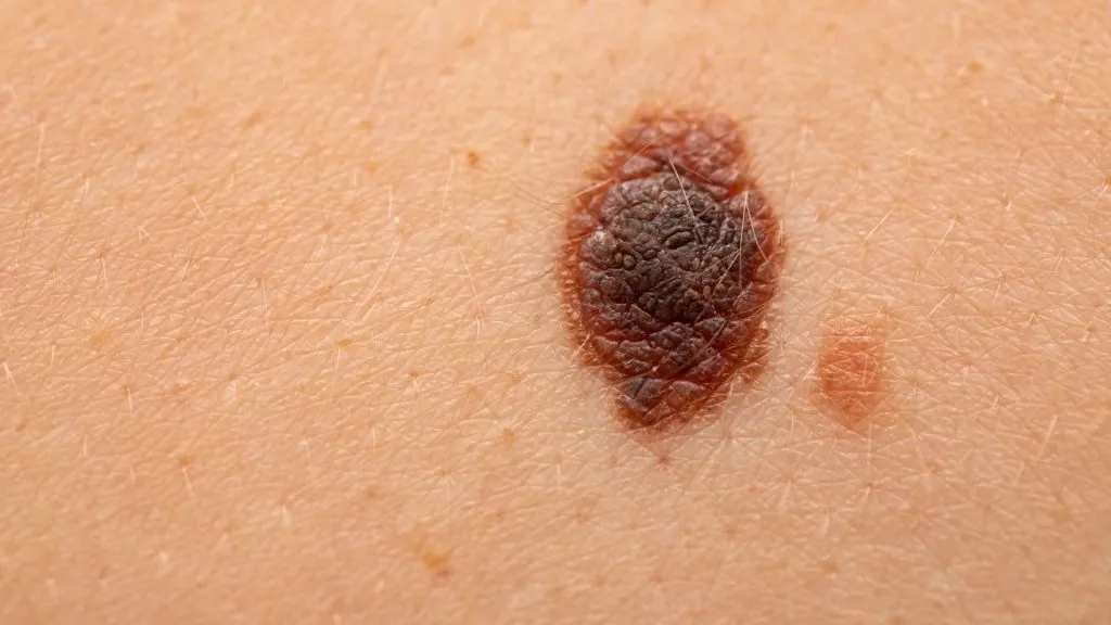
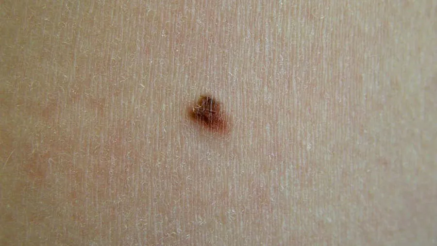
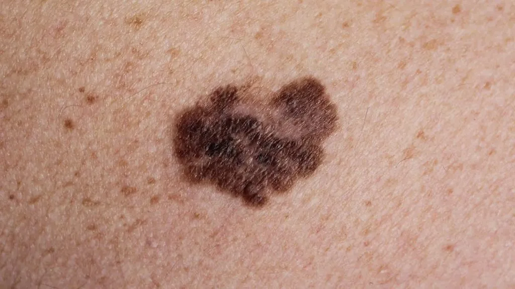
Early melanoma often has those appearances, such as a new pigmented patch on the skin or an unusal change in a pre-existing nevus. They can be primarily identified by the ABCED rule: Asymmetry, Border, Colour, Diameter over 6 mm, Evolution in a short period of time. Patients may have ulcers,bleeding or enlarged surrounding lymph nodes in the advanced stages. Cancer cells will be easy to spread to organs through the blood or lymphatic system without timely treatment, such as lungs,liver and brain. It will cause deterioration of prognosis. At present,surgical resection is the main treatment. There are also some assistant ways: immunotherapy,targeted drugs and radiotherapy. The five-year survival rate for early-stage patients is more than 90%, while the late-stage is less than 25%. We recommended high-risk groups to accept annual dermatological specialist examinations, protect themselves from daily sun and pay attention to normal skin changes.
How is melanoma induced?
Genetic and environmental factors all lead to melanoma. Ultraviolet light exposure is one of the environmental factors,especially intermittent high-intensity sun exposure. Because sunburn will directly damage melanocyte DNA and result in gene mutations. We also should not ignore genetic factors. About 10% of patients have a family history of the disease. According to the research, specific gene mutation increase the risk. In addition, People who has lighter skin colour, multiple atypical nevi or congenital giant nevi are easier to become the patients. Because their skin have weaker melanin protection or more active nevus cells. Abnormal immune system, for example, long-period use of immunosuppressive drugs after organ transplantation, may weaken the body’s ability to remove cancerous cells. Chemical carcinogen exposure or long-term chronic friction irritation are also found as the factors of promoting lesions in recent studies. Not only sunlight but also artificial UV devices provide UV rays, which is dangerous. Some protective ways are recommended: avoid exposure to sunlight, do good physical sun protection, screen regularly for high-risk groups,and take early detection. These methods have a very significant effect in increasing the cure rate.
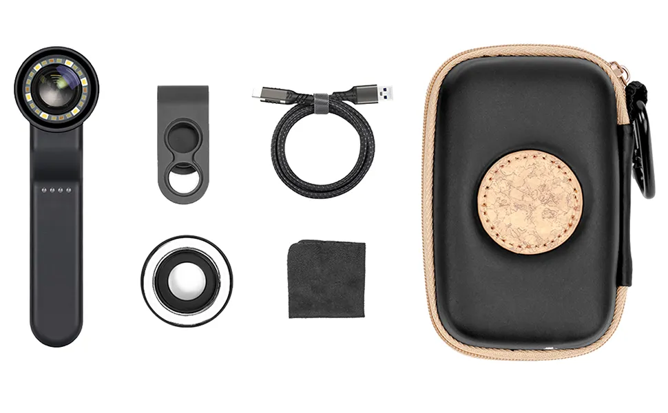
How to conduct a melanoma examination with IBOOLO’s dermatoscope?
IBOOLO’s dermoscopy is an effective technique used in early screening of melanoma, which is non-invasive. It use polarised light to elimate reflections on the skin surface, helping the doctors to clearly observe the complete structures from the epidermis to the superficial dermis. High-resolution images of pigmented lesions will be captured by the magnifying lens of the dermatoscope during the examination. Doctors can analyse features such as colour distribution,blood vessel morphology, pigment network, structural symmetry and so on. For example, irregular grid breaks, blue-white curtain-like structures, or heterogeneous blood vessels are features of malignant melanomas. While benign nevi show as a homogeneous pattern.Compared with visual observation, IBOOLO’s DE-4100 and other series of dermatoscopes can increase the diagnostic accuracy of melanoma by 20-30%. It especially has excellent achievement in determining skin lesions with blurred borders or complex colours. However, we should be careful that dermoscopic results need to be combined with clinical experience and pathological biopsy. Only by this way can we get a right judgement. For this reason, we advice that high-risk individuals check themselves annually and accept timely intervention.
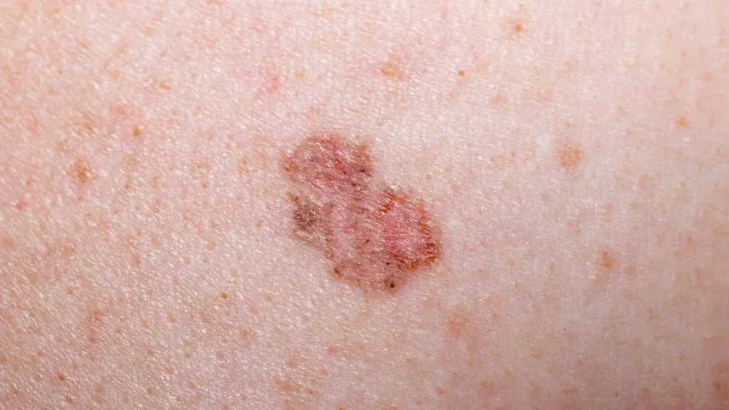
What are the characteristics of melanoma under the IBOOLO skin microscope?
When we observe with iboolo‘s dermatoscope, it is easy to recognise a melanoma based on the following features. Because they are significantly different from benign nevus. One of the important sign is the irregular pigmentation network, it appears as the fracture, thickening or localised disappearance of a dark brown grid, which are similar to snapped branches. In the center of lesion, it exists a blue-grey patch overlying a white structureless area, which is called a blue-white curtain-like structure. This appearance reveals the tumour cells have invaded the dermis. Additionally, malignant lesions are often accompanied by heterogeneous vessels and polychromatic distribution. Heterogeneous vessels are dotted, hairpin or serpentine. The polychromatic distribution are mixed by black, brown, red and blue colours. Other high-risk features include loss of radial striations, negative pigment networks, and degenerative structures. There are someting should be remember:we had better keep high attention on asymmetry, blurred borders, mixed colours, and structural diversity, which are viewed as malignant potential. Although dermoscopy can increase the diagnostic accuracy to more than 80%, it is still necessary to confirm the diagnosis with pathological biopsy. Check skin regularly with dermoscopy can help find changes in the lesions and intervent earlier.
How to prevent melanoma?
Preventing melanoma requires both reducing risk factors. First and foremost, we should take measures for strict sun protection. From 10:00 to 16:00, we will meet the strongest UV rays. So we had better avoid exposing during that time. When we go out, it is crucial to wear a wide-brimmed hat, sun-protective clothing and UV sunglasses. Smearing ourselves with SPF 30 or higher sunscreen is also important. All of the methods should also be sticked on cloudy days or in winter.